7QA4

Crystal structure of stabilized H3N2 A/Hong Kong/1/1968 Hemagglutinin at 2.2 Angstrom


Experimental Data Snapshot

  • Method: X-RAY DIFFRACTION
  • Resolution: 2.19 Å
  • R-Value Free: 0.251 
  • R-Value Work: 0.198 
  • R-Value Observed: 0.201 

wwPDB Validation   3D Report Full Report


Ligand Structure Quality Assessment 


This is version 1.1 of the entry. See complete history


Literature

Universal stabilization of the influenza hemagglutinin by structure-based redesign of the pH switch regions.

Milder, F.J.Jongeneelen, M.Ritschel, T.Bouchier, P.Bisschop, I.J.M.de Man, M.Veldman, D.Le, L.Kaufmann, B.Bakkers, M.J.G.Juraszek, J.Brandenburg, B.Langedijk, J.P.M.

(2022) Proc Natl Acad Sci U S A 119

  • DOI: https://doi.org/10.1073/pnas.2115379119
  • Primary Citation of Related Structures:  
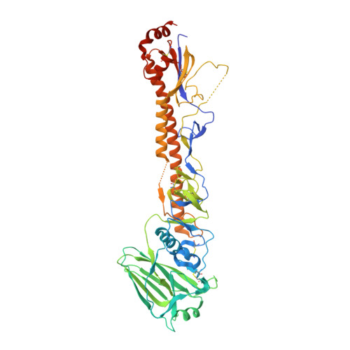
    7QA4

  • PubMed Abstract: 

    For an efficacious vaccine immunogen, influenza hemagglutinin (HA) needs to maintain a stable quaternary structure, which is contrary to the inherently dynamic and metastable nature of class I fusion proteins. In this study, we stabilized HA with three substitutions within its pH-sensitive regions where the refolding starts. An X-ray structure reveals how these substitutions stabilize the intersubunit β-sheet in the base and form an interprotomeric aliphatic layer across the stem while the native prefusion HA fold is retained. The identification of the stabilizing substitutions increases our understanding of how the pH sensitivity is structurally accomplished in HA and possibly other pH-sensitive class I fusion proteins. Our stabilization approach in combination with the occasional back mutation of rare amino acids to consensus results in well-expressing stable trimeric HAs. This repair and stabilization approach, which proves broadly applicable to all tested influenza A HAs of group 1 and 2, will improve the developability of influenza vaccines based on different types of platforms and formats and can potentially improve efficacy.


  • Organizational Affiliation

    Janssen Vaccines & Prevention BV, 2333 CN Leiden, The Netherlands.


Macromolecules
Find similar proteins by:  (by identity cutoff)  |  3D Structure
Entity ID: 1
MoleculeChains Sequence LengthOrganismDetailsImage
Hemagglutinin512Influenza A virus (A/Hong Kong/1/1968(H3N2))Mutation(s): 0 
Gene Names: HA
UniProt
Find proteins for Q91MA7 (Influenza A virus (strain A/Hong Kong/1/1968 H3N2))
Explore Q91MA7 
Go to UniProtKB:  Q91MA7
Entity Groups  
Sequence Clusters30% Identity50% Identity70% Identity90% Identity95% Identity100% Identity
UniProt GroupQ91MA7
Sequence Annotations
Expand
  • Reference Sequence
Oligosaccharides

Help

Entity ID: 2
MoleculeChains Length2D Diagram Glycosylation3D Interactions
beta-D-mannopyranose-(1-4)-2-acetamido-2-deoxy-beta-D-glucopyranose-(1-4)-2-acetamido-2-deoxy-beta-D-glucopyranose
B
3N-Glycosylation
Glycosylation Resources
GlyTouCan:  G15407YE
GlyCosmos:  G15407YE
GlyGen:  G15407YE
Entity ID: 3
MoleculeChains Length2D Diagram Glycosylation3D Interactions
alpha-D-mannopyranose-(1-3)-[alpha-D-mannopyranose-(1-6)]beta-D-mannopyranose-(1-4)-2-acetamido-2-deoxy-beta-D-glucopyranose-(1-4)-2-acetamido-2-deoxy-beta-D-glucopyranose
C
5N-Glycosylation
Glycosylation Resources
GlyTouCan:  G22768VO
GlyCosmos:  G22768VO
GlyGen:  G22768VO
Entity ID: 4
MoleculeChains Length2D Diagram Glycosylation3D Interactions
2-acetamido-2-deoxy-beta-D-glucopyranose-(1-4)-2-acetamido-2-deoxy-beta-D-glucopyranose
D, E
2N-Glycosylation
Glycosylation Resources
GlyTouCan:  G42666HT
GlyCosmos:  G42666HT
GlyGen:  G42666HT
Small Molecules
Ligands 2 Unique
IDChains Name / Formula / InChI Key2D Diagram3D Interactions
NAG
Query on NAG

Download Ideal Coordinates CCD File 
F [auth A]2-acetamido-2-deoxy-beta-D-glucopyranose
C8 H15 N O6
OVRNDRQMDRJTHS-FMDGEEDCSA-N
NO3
Query on NO3

Download Ideal Coordinates CCD File 
G [auth A]NITRATE ION
N O3
NHNBFGGVMKEFGY-UHFFFAOYSA-N
Experimental Data & Validation

Experimental Data

  • Method: X-RAY DIFFRACTION
  • Resolution: 2.19 Å
  • R-Value Free: 0.251 
  • R-Value Work: 0.198 
  • R-Value Observed: 0.201 
  • Space Group: I 21 3
Unit Cell:
Length ( Å )Angle ( ˚ )
a = 154.094α = 90
b = 154.094β = 90
c = 154.094γ = 90
Software Package:
Software NamePurpose
XDSdata reduction
XSCALEdata scaling
REFMACrefinement
PDB_EXTRACTdata extraction
PHASERphasing

Structure Validation

View Full Validation Report


Ligand Structure Quality Assessment 


Entry History & Funding Information

Deposition Data


Funding OrganizationLocationGrant Number
Other governmentUnited StatesHHSO100201700018C

Revision History  (Full details and data files)

  • Version 1.0: 2022-02-02
    Type: Initial release
  • Version 1.1: 2022-03-30
    Changes: Database references