7OXH

ttSlyD with pseudo-wild-type S2 peptide


Experimental Data Snapshot

  • Method: X-RAY DIFFRACTION
  • Resolution: 1.70 Å
  • R-Value Free: 0.238 
  • R-Value Work: 0.203 
  • R-Value Observed: 0.204 

wwPDB Validation   3D Report Full Report


This is version 1.1 of the entry. See complete history


Literature

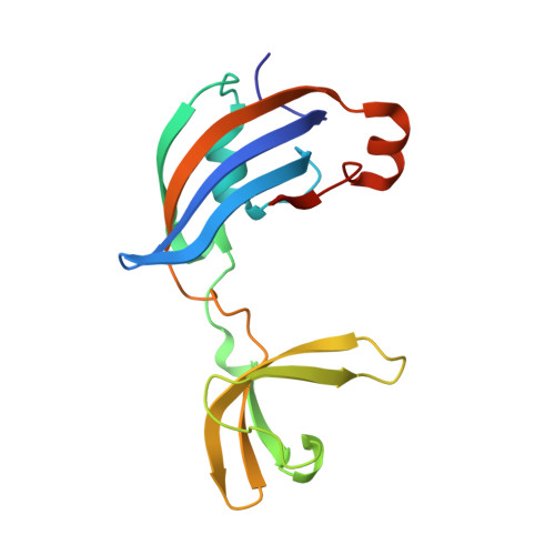
Impact of distant peptide substrate residues on enzymatic activity of SlyD.

Pazicky, S.Werle, A.A.Lei, J.Low, C.Weininger, U.

(2022) Cell Mol Life Sci 79: 138-138

  • DOI: https://doi.org/10.1007/s00018-022-04179-4
  • Primary Citation of Related Structures:  
    7OXG, 7OXH, 7OXI, 7OXJ, 7OXK

  • PubMed Abstract: 

    Peptidyl-prolyl isomerases (PPIases) catalyze intrinsically slow and often rate-limiting isomerization of prolyl-peptide bonds in unfolded or partially folded proteins, thereby speeding up the folding process and preventing misfolding. They often possess binding and chaperone domains in addition to the domain carrying the isomerization activity. Although generally, their substrates display no identity in their amino acid sequence upstream and downstream of the proline with 20 possibilities for each residue, PPIases are efficient enzymes. SlyD is a highly efficient PPIase consisting of an isomerase domain and an additional chaperone domain. The binding of peptide substrates to SlyD and its enzymatic activity depend to some extend on the proline-proximal residues, however, the impact of proline-distant residues has not been investigated so far. Here, we introduce a label-free NMR-based method to measure SlyD activity on different peptide substrates and analysed the data in the context of obtained binding affinities and several co-crystal structures. We show that especially charged and aromatic residues up to eight positions downstream and three positions upstream of the proline and outside the canonical region of similar conformations affect the activity and binding, although they rarely display distinct conformations in our crystal structures. We hypothesize that these positions primarily influence the association reaction. In the absence of the chaperone domain the isomerase activity strongly correlates with substrate affinity, whereas additional factors play a role in its presence. The mutual orientation of isomerase and chaperone domains depends on the presence of substrates in both binding sites, implying allosteric regulation of enzymatic activity.


  • Organizational Affiliation

    Centre for Structural Systems Biology (CSSB), Notkestrasse 85, 22607, Hamburg, Germany.


Macromolecules
Find similar proteins by:  (by identity cutoff)  |  3D Structure
Entity ID: 1
MoleculeChains Sequence LengthOrganismDetailsImage
Peptidyl-prolyl cis-trans isomerase158Thermus thermophilus HB8Mutation(s): 0 
Gene Names: TTHA0346
EC: 5.2.1.8
UniProt
Find proteins for Q5SLE7 (Thermus thermophilus (strain ATCC 27634 / DSM 579 / HB8))
Explore Q5SLE7 
Go to UniProtKB:  Q5SLE7
Entity Groups  
Sequence Clusters30% Identity50% Identity70% Identity90% Identity95% Identity100% Identity
UniProt GroupQ5SLE7
Sequence Annotations
Expand
  • Reference Sequence

Find similar proteins by:  Sequence   |   3D Structure  

Entity ID: 2
MoleculeChains Sequence LengthOrganismDetailsImage
30S ribosomal protein S215Escherichia coli K-12Mutation(s): 3 
UniProt
Find proteins for P0A7V0 (Escherichia coli (strain K12))
Explore P0A7V0 
Go to UniProtKB:  P0A7V0
Entity Groups  
Sequence Clusters30% Identity50% Identity70% Identity90% Identity95% Identity100% Identity
UniProt GroupP0A7V0
Sequence Annotations
Expand
  • Reference Sequence

Find similar proteins by:  Sequence   |   3D Structure  

Entity ID: 3
MoleculeChains Sequence LengthOrganismDetailsImage
Fragment of 30S ribosomal protein S2 peptideC [auth E]2synthetic constructMutation(s): 0 
Sequence Annotations
Expand
  • Reference Sequence
Small Molecules
Ligands 4 Unique
IDChains Name / Formula / InChI Key2D Diagram3D Interactions
PE4
Query on PE4

Download Ideal Coordinates CCD File 
D [auth A],
E [auth A],
G [auth A]
2-{2-[2-(2-{2-[2-(2-ETHOXY-ETHOXY)-ETHOXY]-ETHOXY}-ETHOXY)-ETHOXY]-ETHOXY}-ETHANOL
C16 H34 O8
PJWQOENWHPEPKI-UHFFFAOYSA-N
PEG
Query on PEG

Download Ideal Coordinates CCD File 
F [auth A]DI(HYDROXYETHYL)ETHER
C4 H10 O3
MTHSVFCYNBDYFN-UHFFFAOYSA-N
NI
Query on NI

Download Ideal Coordinates CCD File 
H [auth A],
I [auth A]
NICKEL (II) ION
Ni
VEQPNABPJHWNSG-UHFFFAOYSA-N
CL
Query on CL

Download Ideal Coordinates CCD File 
J [auth A]CHLORIDE ION
Cl
VEXZGXHMUGYJMC-UHFFFAOYSA-M
Experimental Data & Validation

Experimental Data

  • Method: X-RAY DIFFRACTION
  • Resolution: 1.70 Å
  • R-Value Free: 0.238 
  • R-Value Work: 0.203 
  • R-Value Observed: 0.204 
  • Space Group: P 31 2 1
Unit Cell:
Length ( Å )Angle ( ˚ )
a = 49.225α = 90
b = 49.225β = 90
c = 131.214γ = 120
Software Package:
Software NamePurpose
PHENIXrefinement
PHASERphasing
Aimlessdata scaling
XDSdata reduction

Structure Validation

View Full Validation Report


Entry History & Funding Information

Deposition Data


Funding OrganizationLocationGrant Number
Joachim Herz StiftungGermany800026
German Federal Ministry for Education and ResearchGermany05K18YEA

Revision History  (Full details and data files)

  • Version 1.0: 2022-03-16
    Type: Initial release
  • Version 1.1: 2024-01-31
    Changes: Data collection, Refinement description