7ORR

Non-structural protein 10 (nsp10) from SARS CoV-2 in complex with fragment VT00022


Experimental Data Snapshot

  • Method: X-RAY DIFFRACTION
  • Resolution: 1.79 Å
  • R-Value Free: 0.184 
  • R-Value Work: 0.172 
  • R-Value Observed: 0.172 

wwPDB Validation   3D Report Full Report


Ligand Structure Quality Assessment 


This is version 1.2 of the entry. See complete history


Literature

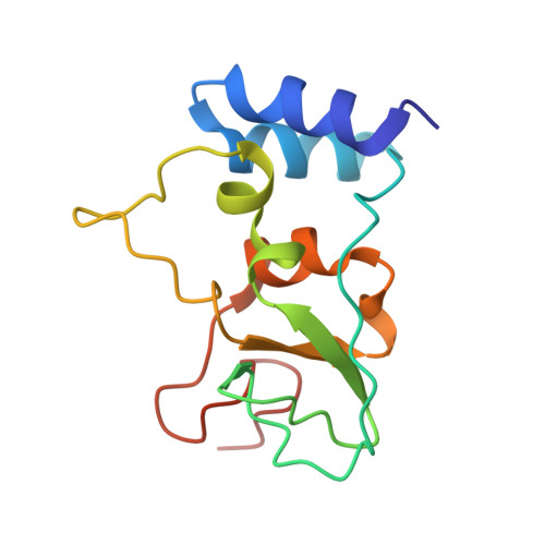
Identification of fragments binding to SARS-CoV-2 nsp10 reveals ligand-binding sites in conserved interfaces between nsp10 and nsp14/nsp16.

Kozielski, F.Sele, C.Talibov, V.O.Lou, J.Dong, D.Wang, Q.Shi, X.Nyblom, M.Rogstam, A.Krojer, T.Fisher, Z.Knecht, W.

(2022) RSC Chem Biol 3: 44-55

  • DOI: https://doi.org/10.1039/d1cb00135c
  • Primary Citation of Related Structures:  
    7ORR, 7ORU, 7ORV, 7ORW

  • PubMed Abstract: 

    Since the emergence of SARS-CoV-2 in 2019, Covid-19 has developed into a serious threat to our health, social and economic systems. Although vaccines have been developed in a tour-de-force and are now increasingly available, repurposing of existing drugs has been less successful. There is a clear need to develop new drugs against SARS-CoV-2 that can also be used against future coronavirus infections. Non-structural protein 10 (nsp10) is a conserved stimulator of two enzymes crucial for viral replication, nsp14 and nsp16, exhibiting exoribonuclease and methyltransferase activities. Interfering with RNA proofreading or RNA cap formation represents intervention strategies to inhibit replication. We applied fragment-based screening using nano differential scanning fluorometry and X-ray crystallography to identify ligands targeting SARS-CoV-2 nsp10. We identified four fragments located in two distinct sites: one can be modelled to where it would be located in the nsp14-nsp10 complex interface and the other in the nsp16-nsp10 complex interface. Microscale thermophoresis (MST) experiments were used to quantify fragment affinities for nsp10. Additionally, we showed by MST that the interaction by nsp14 and 10 is weak and thereby that complex formation could be disrupted by small molecules. The fragments will serve as starting points for the development of more potent analogues using fragment growing techniques and structure-based drug design.


  • Organizational Affiliation

    School of Pharmacy, University College London 29-39 Brunswick Square London WC1N 1AX UK.


Macromolecules
Find similar proteins by:  (by identity cutoff)  |  3D Structure
Entity ID: 1
MoleculeChains Sequence LengthOrganismDetailsImage
Non-structural protein 10125Severe acute respiratory syndrome coronavirus 2Mutation(s): 0 
Gene Names: rep1a-1b
UniProt
Find proteins for P0DTD1 (Severe acute respiratory syndrome coronavirus 2)
Explore P0DTD1 
Go to UniProtKB:  P0DTD1
Entity Groups  
Sequence Clusters30% Identity50% Identity70% Identity90% Identity95% Identity100% Identity
UniProt GroupP0DTD1
Sequence Annotations
Expand
  • Reference Sequence
Small Molecules
Ligands 5 Unique
IDChains Name / Formula / InChI Key2D Diagram3D Interactions
PIM (Subject of Investigation/LOI)
Query on PIM

Download Ideal Coordinates CCD File 
F [auth A],
G [auth A]
4-PHENYL-1H-IMIDAZOLE
C9 H8 N2
XHLKOHSAWQPOFO-UHFFFAOYSA-N
GOL
Query on GOL

Download Ideal Coordinates CCD File 
H [auth A],
I [auth A]
GLYCEROL
C3 H8 O3
PEDCQBHIVMGVHV-UHFFFAOYSA-N
DMS
Query on DMS

Download Ideal Coordinates CCD File 
D [auth A],
E [auth A]
DIMETHYL SULFOXIDE
C2 H6 O S
IAZDPXIOMUYVGZ-UHFFFAOYSA-N
ZN
Query on ZN

Download Ideal Coordinates CCD File 
B [auth A],
C [auth A]
ZINC ION
Zn
PTFCDOFLOPIGGS-UHFFFAOYSA-N
CL
Query on CL

Download Ideal Coordinates CCD File 
J [auth A],
K [auth A]
CHLORIDE ION
Cl
VEXZGXHMUGYJMC-UHFFFAOYSA-M
Experimental Data & Validation

Experimental Data

  • Method: X-RAY DIFFRACTION
  • Resolution: 1.79 Å
  • R-Value Free: 0.184 
  • R-Value Work: 0.172 
  • R-Value Observed: 0.172 
  • Space Group: I 21 3
  • Diffraction Data: https://doi.org/10.18430/M37ORR
Unit Cell:
Length ( Å )Angle ( ˚ )
a = 105.883α = 90
b = 105.883β = 90
c = 105.883γ = 90
Software Package:
Software NamePurpose
Aimlessdata scaling
REFMACrefinement
PDB_EXTRACTdata extraction
XDSdata reduction
PHASERphasing

Structure Validation

View Full Validation Report


Ligand Structure Quality Assessment 


Entry History & Funding Information

Deposition Data


Funding OrganizationLocationGrant Number
Swedish Research CouncilSweden2018-06454
Medical Research Council (MRC, United Kingdom)United KingdomCiC8 2019 MC_PC_19054

Revision History  (Full details and data files)

  • Version 1.0: 2021-10-20
    Type: Initial release
  • Version 1.1: 2022-02-23
    Changes: Database references
  • Version 1.2: 2024-01-31
    Changes: Data collection, Refinement description