7OFH

CryoEM structure of the outer membrane secretin pore pIV from the f1 filamentous bacteriophage.


Experimental Data Snapshot

  • Method: ELECTRON MICROSCOPY
  • Resolution: 2.70 Å
  • Aggregation State: PARTICLE 
  • Reconstruction Method: SINGLE PARTICLE 

wwPDB Validation   3D Report Full Report


This is version 1.1 of the entry. See complete history


Literature

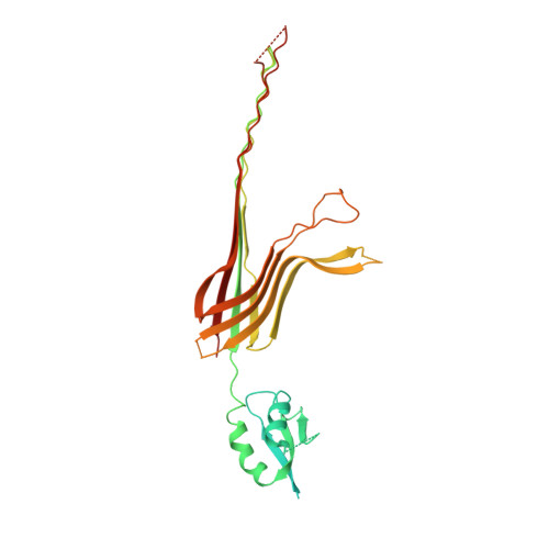
CryoEM structure of the outer membrane secretin channel pIV from the f1 filamentous bacteriophage.

Conners, R.McLaren, M.Lapinska, U.Sanders, K.Stone, M.R.L.Blaskovich, M.A.T.Pagliara, S.Daum, B.Rakonjac, J.Gold, V.A.M.

(2021) Nat Commun 12: 6316-6316

  • DOI: https://doi.org/10.1038/s41467-021-26610-3
  • Primary Citation of Related Structures:  
    7OFH

  • PubMed Abstract: 

    The Ff family of filamentous bacteriophages infect gram-negative bacteria, but do not cause lysis of their host cell. Instead, new virions are extruded via the phage-encoded pIV protein, which has homology with bacterial secretins. Here, we determine the structure of pIV from the f1 filamentous bacteriophage at 2.7 Å resolution by cryo-electron microscopy, the first near-atomic structure of a phage secretin. Fifteen f1 pIV subunits assemble to form a gated channel in the bacterial outer membrane, with associated soluble domains projecting into the periplasm. We model channel opening and propose a mechanism for phage egress. By single-cell microfluidics experiments, we demonstrate the potential for secretins such as pIV to be used as adjuvants to increase the uptake and efficacy of antibiotics in bacteria. Finally, we compare the f1 pIV structure to its homologues to reveal similarities and differences between phage and bacterial secretins.


  • Organizational Affiliation

    Living Systems Institute, University of Exeter, Exeter, UK.


Macromolecules
Find similar proteins by:  (by identity cutoff)  |  3D Structure
Entity ID: 1
MoleculeChains Sequence LengthOrganismDetailsImage
Virion export protein
A, B, C, D, E
A, B, C, D, E, F, G, H, I, J, K, L, M, N, O
414Enterobacteria phage f1Mutation(s): 4 
Gene Names: IV
Membrane Entity: Yes 
UniProt
Find proteins for P03666 (Enterobacteria phage f1)
Explore P03666 
Go to UniProtKB:  P03666
Entity Groups  
Sequence Clusters30% Identity50% Identity70% Identity90% Identity95% Identity100% Identity
UniProt GroupP03666
Sequence Annotations
Expand
  • Reference Sequence
Small Molecules
Ligands 1 Unique
IDChains Name / Formula / InChI Key2D Diagram3D Interactions
CPS
Query on CPS

Download Ideal Coordinates CCD File 
AA [auth F]
BA [auth G]
CA [auth G]
DA [auth H]
EA [auth H]
AA [auth F],
BA [auth G],
CA [auth G],
DA [auth H],
EA [auth H],
FA [auth I],
GA [auth I],
HA [auth J],
IA [auth J],
JA [auth K],
KA [auth K],
LA [auth L],
MA [auth L],
NA [auth M],
OA [auth M],
P [auth A],
PA [auth N],
Q [auth A],
QA [auth N],
R [auth B],
RA [auth O],
S [auth B],
SA [auth O],
T [auth C],
U [auth C],
V [auth D],
W [auth D],
X [auth E],
Y [auth E],
Z [auth F]
3-[(3-CHOLAMIDOPROPYL)DIMETHYLAMMONIO]-1-PROPANESULFONATE
C32 H58 N2 O7 S
UMCMPZBLKLEWAF-BCTGSCMUSA-N
Experimental Data & Validation

Experimental Data

  • Method: ELECTRON MICROSCOPY
  • Resolution: 2.70 Å
  • Aggregation State: PARTICLE 
  • Reconstruction Method: SINGLE PARTICLE 
EM Software:
TaskSoftware PackageVersion
RECONSTRUCTIONRELION3.1beta
MODEL REFINEMENTREFMAC

Structure Validation

View Full Validation Report


Entry History & Funding Information

Deposition Data


Funding OrganizationLocationGrant Number
Wellcome TrustUnited Kingdom210363/Z/18/Z

Revision History  (Full details and data files)

  • Version 1.0: 2021-11-03
    Type: Initial release
  • Version 1.1: 2021-11-17
    Changes: Data collection, Database references