7N39

Crystal structure of 7-site deamidated variant of human gamma(S)-crystallin


Experimental Data Snapshot

  • Method: X-RAY DIFFRACTION
  • Resolution: 1.56 Å
  • R-Value Free: 0.213 
  • R-Value Work: 0.193 
  • R-Value Observed: 0.194 

wwPDB Validation   3D Report Full Report


This is version 1.2 of the entry. See complete history


Literature

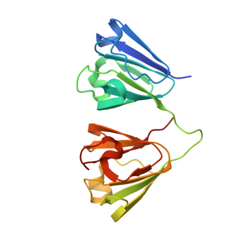
Deamidation of the human eye lens protein gamma S-crystallin accelerates oxidative aging.

Norton-Baker, B.Mehrabi, P.Kwok, A.O.Roskamp, K.W.Rocha, M.A.Sprague-Piercy, M.A.von Stetten, D.Miller, R.J.D.Martin, R.W.

(2022) Structure 30: 763-776.e4

  • DOI: https://doi.org/10.1016/j.str.2022.03.002
  • Primary Citation of Related Structures:  
    7N36, 7N37, 7N38, 7N39, 7N3A, 7N3B

  • PubMed Abstract: 

    Cataract, a clouding of the eye lens from protein precipitation, affects millions of people every year. The lens proteins, the crystallins, show extensive post-translational modifications (PTMs) in cataractous lenses. The most common PTMs, deamidation and oxidation, promote crystallin aggregation; however, it is not clear precisely how these PTMs contribute to crystallin insolubilization. Here, we report six crystal structures of the lens protein γS-crystallin (γS): one of the wild-type and five of deamidated γS variants, from three to nine deamidation sites, after sample aging. The deamidation mutations do not change the overall fold of γS; however, increasing deamidation leads to accelerated disulfide-bond formation. Addition of deamidated sites progressively destabilized protein structure, and the deamidated variants display an increased propensity for aggregation. These results suggest that the deamidated variants are useful as models for accelerated aging; the structural changes observed provide support for redox activity of γS-crystallin in the lens.


  • Organizational Affiliation

    Department of Chemistry, University of California, Irvine, CA 92697-2025, USA; Department for Atomically Resolved Dynamics, Max-Planck-Institute for Structure and Dynamics of Matter, Luruper Chaussee 149, 22761 Hamburg, Germany.


Macromolecules
Find similar proteins by:  (by identity cutoff)  |  3D Structure
Entity ID: 1
MoleculeChains Sequence LengthOrganismDetailsImage
Gamma-crystallin S
A, B
178Homo sapiensMutation(s): 7 
Gene Names: CRYGSCRYG8
UniProt & NIH Common Fund Data Resources
Find proteins for P22914 (Homo sapiens)
Explore P22914 
Go to UniProtKB:  P22914
PHAROS:  P22914
GTEx:  ENSG00000213139 
Entity Groups  
Sequence Clusters30% Identity50% Identity70% Identity90% Identity95% Identity100% Identity
UniProt GroupP22914
Sequence Annotations
Expand
  • Reference Sequence
Small Molecules
Ligands 1 Unique
IDChains Name / Formula / InChI Key2D Diagram3D Interactions
SO4
Query on SO4

Download Ideal Coordinates CCD File 
C [auth A]SULFATE ION
O4 S
QAOWNCQODCNURD-UHFFFAOYSA-L
Experimental Data & Validation

Experimental Data

  • Method: X-RAY DIFFRACTION
  • Resolution: 1.56 Å
  • R-Value Free: 0.213 
  • R-Value Work: 0.193 
  • R-Value Observed: 0.194 
  • Space Group: P 2 21 21
Unit Cell:
Length ( Å )Angle ( ˚ )
a = 49.146α = 90
b = 76.629β = 90
c = 94.655γ = 90
Software Package:
Software NamePurpose
PHENIXrefinement
XDSdata reduction
XSCALEdata scaling
PHASERphasing

Structure Validation

View Full Validation Report


Entry History 

Deposition Data

Revision History  (Full details and data files)

  • Version 1.0: 2022-03-23
    Type: Initial release
  • Version 1.1: 2022-10-05
    Changes: Database references
  • Version 1.2: 2023-10-18
    Changes: Data collection, Refinement description