


Urea binding to guide rational design of mutations that influence enzyme dynamics
Hamill, C.J., Arcus, V.L., Prentice, E.J., Bahl, C., Truebridge, I.To be published.
Experimental Data Snapshot
wwPDB Validation 3D Report Full Report

Entity ID: 1 | |||||
|---|---|---|---|---|---|
| Molecule | Chains | Sequence Length | Organism | Details | Image |
| Oligo-1,6-glucosidase 1 | A [auth B] | 586 | Bacillus subtilis subsp. subtilis str. 168 | Mutation(s): 1 Gene Names: malL, yvdL, BSU34560 EC: 3.2.1.10 | 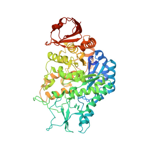 |
UniProt | |||||
Find proteins for O06994 (Bacillus subtilis (strain 168)) Explore O06994 Go to UniProtKB: O06994 | |||||
Entity Groups | |||||
| Sequence Clusters | 30% Identity50% Identity70% Identity90% Identity95% Identity100% Identity | ||||
| UniProt Group | O06994 | ||||
Sequence AnnotationsExpand | |||||
| |||||
| Ligands 3 Unique | |||||
|---|---|---|---|---|---|
| ID | Chains | Name / Formula / InChI Key | 2D Diagram | 3D Interactions | |
| TRS Query on TRS | B | 2-AMINO-2-HYDROXYMETHYL-PROPANE-1,3-DIOL C4 H12 N O3 LENZDBCJOHFCAS-UHFFFAOYSA-O |  | ||
| GOL Query on GOL | C [auth B] | GLYCEROL C3 H8 O3 PEDCQBHIVMGVHV-UHFFFAOYSA-N |  | ||
| CA Query on CA | D [auth B] | CALCIUM ION Ca BHPQYMZQTOCNFJ-UHFFFAOYSA-N |  | ||
| Length ( Å ) | Angle ( ˚ ) |
|---|---|
| a = 48.749 | α = 90 |
| b = 100.999 | β = 113.06 |
| c = 61.749 | γ = 90 |
| Software Name | Purpose |
|---|---|
| XDS | data reduction |
| Aimless | data scaling |
| MOLREP | phasing |
| REFMAC | refinement |
| PHENIX | refinement |
| Coot | model building |
| PDB_EXTRACT | data extraction |
| Funding Organization | Location | Grant Number |
|---|---|---|
| Marsden Fund | New Zealand | 16-UOW-027 |
RCSB PDB (citation) is hosted by
RCSB PDB is a member of the

