7KMB

ACE2-RBD Focused Refinement Using Symmetry Expansion of Applied C3 for Triple ACE2-bound SARS-CoV-2 Trimer Spike at pH 7.4


Experimental Data Snapshot

  • Method: ELECTRON MICROSCOPY
  • Resolution: 3.39 Å
  • Aggregation State: PARTICLE 
  • Reconstruction Method: SINGLE PARTICLE 

wwPDB Validation   3D Report Full Report


This is version 1.3 of the entry. See complete history


Literature

Cryo-EM Structures of SARS-CoV-2 Spike without and with ACE2 Reveal a pH-Dependent Switch to Mediate Endosomal Positioning of Receptor-Binding Domains.

Zhou, T.Tsybovsky, Y.Gorman, J.Rapp, M.Cerutti, G.Chuang, G.Y.Katsamba, P.S.Sampson, J.M.Schon, A.Bimela, J.Boyington, J.C.Nazzari, A.Olia, A.S.Shi, W.Sastry, M.Stephens, T.Stuckey, J.Teng, I.T.Wang, P.Wang, S.Zhang, B.Friesner, R.A.Ho, D.D.Mascola, J.R.Shapiro, L.Kwong, P.D.

(2020) Cell Host Microbe 28: 867-879.e5

  • DOI: https://doi.org/10.1016/j.chom.2020.11.004
  • Primary Citation of Related Structures:  
    6XLU, 6XM0, 6XM3, 6XM4, 6XM5, 7JWY, 7KMB, 7KMS, 7KMZ, 7KNB, 7KNE, 7KNH, 7KNI

  • PubMed Abstract: 

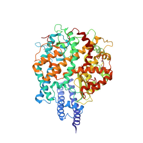
    The SARS-CoV-2 spike employs mobile receptor-binding domains (RBDs) to engage the human ACE2 receptor and to facilitate virus entry, which can occur through low-pH-endosomal pathways. To understand how ACE2 binding and low pH affect spike conformation, we determined cryo-electron microscopy structures-at serological and endosomal pH-delineating spike recognition of up to three ACE2 molecules. RBDs freely adopted "up" conformations required for ACE2 interaction, primarily through RBD movement combined with smaller alterations in neighboring domains. In the absence of ACE2, single-RBD-up conformations dominated at pH 5.5, resolving into a solitary all-down conformation at lower pH. Notably, a pH-dependent refolding region (residues 824-858) at the spike-interdomain interface displayed dramatic structural rearrangements and mediated RBD positioning through coordinated movements of the entire trimer apex. These structures provide a foundation for understanding prefusion-spike mechanics governing endosomal entry; we suggest that the low pH all-down conformation potentially facilitates immune evasion from RBD-up binding antibody.


  • Organizational Affiliation

    Vaccine Research Center, National Institute of Allergy and Infectious Diseases, National Institutes of Health, Bethesda, MD 20892, USA.


Macromolecules
Find similar proteins by:  (by identity cutoff)  |  3D Structure
Entity ID: 1
MoleculeChains Sequence LengthOrganismDetailsImage
Angiotensin-converting enzyme 2A [auth F]597Homo sapiensMutation(s): 0 
Gene Names: ACE2UNQ868/PRO1885
EC: 3.4.17.23 (PDB Primary Data), 3.4.17 (PDB Primary Data)
Membrane Entity: Yes 
UniProt & NIH Common Fund Data Resources
Find proteins for Q9BYF1 (Homo sapiens)
Explore Q9BYF1 
Go to UniProtKB:  Q9BYF1
PHAROS:  Q9BYF1
GTEx:  ENSG00000130234 
Entity Groups  
Sequence Clusters30% Identity50% Identity70% Identity90% Identity95% Identity100% Identity
UniProt GroupQ9BYF1
Sequence Annotations
Expand
  • Reference Sequence
Find similar proteins by:  (by identity cutoff)  |  3D Structure
Entity ID: 2
MoleculeChains Sequence LengthOrganismDetailsImage
Spike glycoproteinB [auth G]1,273Severe acute respiratory syndrome coronavirus 2Mutation(s): 0 
Gene Names: S2
Membrane Entity: Yes 
UniProt
Find proteins for P0DTC2 (Severe acute respiratory syndrome coronavirus 2)
Explore P0DTC2 
Go to UniProtKB:  P0DTC2
Entity Groups  
Sequence Clusters30% Identity50% Identity70% Identity90% Identity95% Identity100% Identity
UniProt GroupP0DTC2
Sequence Annotations
Expand
  • Reference Sequence
Oligosaccharides

Help

Entity ID: 3
MoleculeChains Length2D Diagram Glycosylation3D Interactions
2-acetamido-2-deoxy-beta-D-glucopyranose-(1-4)-2-acetamido-2-deoxy-beta-D-glucopyranoseC [auth A],
D [auth B]
2N-Glycosylation
Glycosylation Resources
GlyTouCan:  G42666HT
GlyCosmos:  G42666HT
GlyGen:  G42666HT
Experimental Data & Validation

Experimental Data

  • Method: ELECTRON MICROSCOPY
  • Resolution: 3.39 Å
  • Aggregation State: PARTICLE 
  • Reconstruction Method: SINGLE PARTICLE 
EM Software:
TaskSoftware PackageVersion
RECONSTRUCTIONcryoSPARC2.15
MODEL REFINEMENTPHENIX

Structure Validation

View Full Validation Report


Entry History & Funding Information

Deposition Data


Funding OrganizationLocationGrant Number
National Institutes of Health/National Institute of General Medical Sciences (NIH/NIGMS)United StatesU24 GM129539
Other privateUnited StatesSF349247 (Simons Foundation)

Revision History  (Full details and data files)

  • Version 1.0: 2020-12-09
    Type: Initial release
  • Version 1.1: 2020-12-16
    Changes: Database references
  • Version 1.2: 2020-12-23
    Changes: Database references
  • Version 1.3: 2021-12-15
    Changes: Database references