7FDH

SARS-COV-2 Spike RBDMACSp25 binding to hACE2


Experimental Data Snapshot

  • Method: ELECTRON MICROSCOPY
  • Resolution: 3.72 Å
  • Aggregation State: PARTICLE 
  • Reconstruction Method: SINGLE PARTICLE 

wwPDB Validation   3D Report Full Report


This is version 1.1 of the entry. See complete history


Literature

Characterization and structural basis of a lethal mouse-adapted SARS-CoV-2.

Sun, S.Gu, H.Cao, L.Chen, Q.Ye, Q.Yang, G.Li, R.T.Fan, H.Deng, Y.Q.Song, X.Qi, Y.Li, M.Lan, J.Feng, R.Guo, Y.Zhu, N.Qin, S.Wang, L.Zhang, Y.F.Zhou, C.Zhao, L.Chen, Y.Shen, M.Cui, Y.Yang, X.Wang, X.Tan, W.Wang, H.Wang, X.Qin, C.F.

(2021) Nat Commun 12: 5654-5654

  • DOI: https://doi.org/10.1038/s41467-021-25903-x
  • Primary Citation of Related Structures:  
    7FDG, 7FDH, 7FDI, 7FDK

  • PubMed Abstract: 

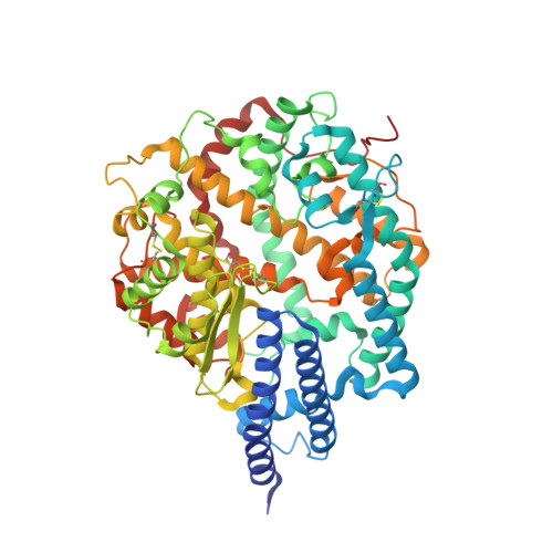
    There is an urgent need for animal models to study SARS-CoV-2 pathogenicity. Here, we generate and characterize a novel mouse-adapted SARS-CoV-2 strain, MASCp36, that causes severe respiratory symptoms, and mortality. Our model exhibits age- and gender-related mortality akin to severe COVID-19. Deep sequencing identified three amino acid substitutions, N501Y, Q493H, and K417N, at the receptor binding domain (RBD) of MASCp36, during in vivo passaging. All three RBD mutations significantly enhance binding affinity to its endogenous receptor, ACE2. Cryo-electron microscopy analysis of human ACE2 (hACE2), or mouse ACE2 (mACE2), in complex with the RBD of MASCp36, at 3.1 to 3.7 Å resolution, reveals the molecular basis for the receptor-binding switch. N501Y and Q493H enhance the binding affinity to hACE2, whereas triple mutations at N501Y/Q493H/K417N decrease affinity and reduce infectivity of MASCp36. Our study provides a platform for studying SARS-CoV-2 pathogenesis, and unveils the molecular mechanism for its rapid adaptation and evolution.


  • Organizational Affiliation

    State Key Laboratory of Pathogen and Biosecurity, Beijing Institute of Microbiology and Epidemiology, AMMS, Beijing, 100071, China.


Macromolecules
Find similar proteins by:  (by identity cutoff)  |  3D Structure
Entity ID: 1
MoleculeChains Sequence LengthOrganismDetailsImage
Angiotensin-converting enzyme 2621Homo sapiensMutation(s): 0 
Gene Names: ACE2UNQ868/PRO1885
EC: 3.4.17.23 (PDB Primary Data), 3.4.17 (PDB Primary Data)
UniProt & NIH Common Fund Data Resources
Find proteins for Q9BYF1 (Homo sapiens)
Explore Q9BYF1 
Go to UniProtKB:  Q9BYF1
PHAROS:  Q9BYF1
GTEx:  ENSG00000130234 
Entity Groups  
Sequence Clusters30% Identity50% Identity70% Identity90% Identity95% Identity100% Identity
UniProt GroupQ9BYF1
Sequence Annotations
Expand
  • Reference Sequence
Find similar proteins by:  (by identity cutoff)  |  3D Structure
Entity ID: 2
MoleculeChains Sequence LengthOrganismDetailsImage
Spike protein S1B [auth E]213Severe acute respiratory syndrome coronavirus 2Mutation(s): 2 
Gene Names: S2
UniProt
Find proteins for P0DTC2 (Severe acute respiratory syndrome coronavirus 2)
Explore P0DTC2 
Go to UniProtKB:  P0DTC2
Entity Groups  
Sequence Clusters30% Identity50% Identity70% Identity90% Identity95% Identity100% Identity
UniProt GroupP0DTC2
Sequence Annotations
Expand
  • Reference Sequence
Experimental Data & Validation

Experimental Data

  • Method: ELECTRON MICROSCOPY
  • Resolution: 3.72 Å
  • Aggregation State: PARTICLE 
  • Reconstruction Method: SINGLE PARTICLE 

Structure Validation

View Full Validation Report


Entry History 

Deposition Data

Revision History  (Full details and data files)

  • Version 1.0: 2021-08-25
    Type: Initial release
  • Version 1.1: 2022-03-23
    Changes: Database references