7E9K

Crystal Structure of POMGNT2 in complex with UDP and mono-mannosyl peptide (379Man long peptide)

  • Classification: TRANSFERASE
  • Organism(s): Bos taurus, Homo sapiens
  • Expression System: Homo sapiens
  • Mutation(s): No 

  • Deposited: 2021-03-04 Released: 2021-05-05 
  • Deposition Author(s): Kuwabara, N.
  • Funding Organization(s): Japan Society for the Promotion of Science (JSPS), Japan Agency for Medical Research and Development (AMED)

Experimental Data Snapshot

  • Method: X-RAY DIFFRACTION
  • Resolution: 2.05 Å
  • R-Value Free: 0.241 
  • R-Value Work: 0.202 
  • R-Value Observed: 0.204 

wwPDB Validation   3D Report Full Report


Ligand Structure Quality Assessment 


This is version 1.2 of the entry. See complete history


Literature

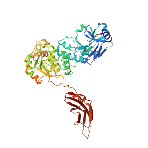
The structure of POMGNT2 provides new insights into the mechanism to determine the functional O-mannosylation site on alpha-dystroglycan.

Imae, R.Kuwabara, N.Manya, H.Tanaka, T.Tsuyuguchi, M.Mizuno, M.Endo, T.Kato, R.

(2021) Genes Cells 26: 485-494

  • DOI: https://doi.org/10.1111/gtc.12853
  • Primary Citation of Related Structures:  
    7E9J, 7E9K, 7E9L

  • PubMed Abstract: 

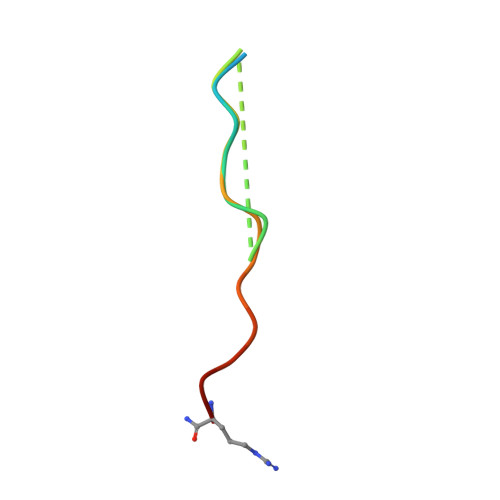
    Defects in the O-mannosyl glycan of α-dystroglycan (α-DG) are associated with α-dystroglycanopathy, a group of congenital muscular dystrophies. While α-DG has many O-mannosylation sites, only the specific positions can be modified with the functional O-mannosyl glycan, namely, core M3-type glycan. POMGNT2 is a glycosyltransferase which adds β1,4-linked GlcNAc to the O-mannose (Man) residue to acquire core M3-type glycan. Although it is assumed that POMGNT2 extends the specific O-Man residues around particular amino acid sequences, the details are not well understood. Here, we determined a series of crystal structures of POMGNT2 with and without the acceptor O-mannosyl peptides and identified the critical interactions between POMGNT2 and the acceptor peptide. POMGNT2 has an N-terminal catalytic domain and a C-terminal fibronectin type III (FnIII) domain and forms a dimer. The acceptor peptide is sandwiched between the two protomers. The catalytic domain of one protomer recognizes the O-mannosylation site (TPT motif), and the FnIII domain of the other protomer recognizes the C-terminal region of the peptide. Structure-based mutational studies confirmed that amino acid residues of the catalytic domain interacting with mannose or the TPT motif are essential for POMGNT2 enzymatic activity. In addition, the FnIII domain is also essential for the activity and it interacts with the peptide mainly by hydrophobic interaction. Our study provides the first atomic-resolution insights into specific acceptor recognition by the FnIII domain of POMGNT2. The catalytic mechanism of POMGNT2 is proposed based on the structure.


  • Organizational Affiliation

    Molecular Glycobiology, Research Team for Mechanism of Aging, Tokyo Metropolitan Geriatric Hospital and Institute of Gerontology, Itabashi-ku, Japan.


Macromolecules
Find similar proteins by:  (by identity cutoff)  |  3D Structure
Entity ID: 1
MoleculeChains Sequence LengthOrganismDetailsImage
Protein O-linked-mannose beta-1,4-N-acetylglucosaminyltransferase 2A,
B,
C [auth D],
D [auth E]
539Bos taurusMutation(s): 0 
Gene Names: POMGNT2AGO61GTDC2
EC: 2.4.1.312
UniProt
Find proteins for Q5NDF2 (Bos taurus)
Explore Q5NDF2 
Go to UniProtKB:  Q5NDF2
Entity Groups  
Sequence Clusters30% Identity50% Identity70% Identity90% Identity95% Identity100% Identity
UniProt GroupQ5NDF2
Sequence Annotations
Expand
  • Reference Sequence

Find similar proteins by:  Sequence   |   3D Structure  

Entity ID: 2
MoleculeChains Sequence LengthOrganismDetailsImage
mono-mannosyl peptide (379Man long peptide)E [auth C],
F
23Homo sapiensMutation(s): 0 
UniProt & NIH Common Fund Data Resources
Find proteins for Q14118 (Homo sapiens)
Explore Q14118 
Go to UniProtKB:  Q14118
PHAROS:  Q14118
GTEx:  ENSG00000173402 
Entity Groups  
Sequence Clusters30% Identity50% Identity70% Identity90% Identity95% Identity100% Identity
UniProt GroupQ14118
Sequence Annotations
Expand
  • Reference Sequence
Oligosaccharides

Help

Entity ID: 3
MoleculeChains Length2D Diagram Glycosylation3D Interactions
2-acetamido-2-deoxy-beta-D-glucopyranose-(1-4)-2-acetamido-2-deoxy-beta-D-glucopyranose
G
2N-Glycosylation
Glycosylation Resources
GlyTouCan:  G42666HT
GlyCosmos:  G42666HT
GlyGen:  G42666HT
Small Molecules
Ligands 4 Unique
IDChains Name / Formula / InChI Key2D Diagram3D Interactions
UDP (Subject of Investigation/LOI)
Query on UDP

Download Ideal Coordinates CCD File 
K [auth A],
Q [auth B],
T [auth D],
Y [auth E]
URIDINE-5'-DIPHOSPHATE
C9 H14 N2 O12 P2
XCCTYIAWTASOJW-XVFCMESISA-N
NAG
Query on NAG

Download Ideal Coordinates CCD File 
H [auth A]
I [auth A]
J [auth A]
M [auth B]
N [auth B]
H [auth A],
I [auth A],
J [auth A],
M [auth B],
N [auth B],
O [auth B],
P [auth B],
R [auth D],
S [auth D],
V [auth E],
W [auth E],
X [auth E]
2-acetamido-2-deoxy-beta-D-glucopyranose
C8 H15 N O6
OVRNDRQMDRJTHS-FMDGEEDCSA-N
MAN (Subject of Investigation/LOI)
Query on MAN

Download Ideal Coordinates CCD File 
AA [auth F],
Z [auth C]
alpha-D-mannopyranose
C6 H12 O6
WQZGKKKJIJFFOK-PQMKYFCFSA-N
TRS (Subject of Investigation/LOI)
Query on TRS

Download Ideal Coordinates CCD File 
L [auth A],
U [auth D]
2-AMINO-2-HYDROXYMETHYL-PROPANE-1,3-DIOL
C4 H12 N O3
LENZDBCJOHFCAS-UHFFFAOYSA-O
Experimental Data & Validation

Experimental Data

  • Method: X-RAY DIFFRACTION
  • Resolution: 2.05 Å
  • R-Value Free: 0.241 
  • R-Value Work: 0.202 
  • R-Value Observed: 0.204 
  • Space Group: P 1 21 1
Unit Cell:
Length ( Å )Angle ( ˚ )
a = 86.15α = 90
b = 104.401β = 90.104
c = 148.639γ = 90
Software Package:
Software NamePurpose
PHENIXrefinement
XDSdata reduction
Aimlessdata scaling
PHASERphasing

Structure Validation

View Full Validation Report


Ligand Structure Quality Assessment 


Entry History & Funding Information

Deposition Data

  • Released Date: 2021-05-05 
  • Deposition Author(s): Kuwabara, N.

Funding OrganizationLocationGrant Number
Japan Society for the Promotion of Science (JSPS)JapanJP16K07284
Japan Agency for Medical Research and Development (AMED)JapanJP20am0101083
Japan Society for the Promotion of Science (JSPS)JapanJP19H05648
Japan Society for the Promotion of Science (JSPS)JapanJP17H03987

Revision History  (Full details and data files)

  • Version 1.0: 2021-05-05
    Type: Initial release
  • Version 1.1: 2021-07-28
    Changes: Database references
  • Version 1.2: 2024-04-03
    Changes: Data collection, Database references, Refinement description