7B7H

The glucuronoyl esterase OtCE15A R268A variant from Opitutus terrae in complex with, and covalently linked to, D-glucuronate


Experimental Data Snapshot

  • Method: X-RAY DIFFRACTION
  • Resolution: 1.80 Å
  • R-Value Free: 0.222 
  • R-Value Work: 0.183 
  • R-Value Observed: 0.185 

wwPDB Validation   3D Report Full Report


Ligand Structure Quality Assessment 


This is version 1.2 of the entry. See complete history


Literature

Mechanism and biomass association of glucuronoyl esterase: an alpha / beta hydrolase with potential in biomass conversion.

Zong, Z.Mazurkewich, S.Pereira, C.S.Fu, H.Cai, W.Shao, X.Skaf, M.S.Larsbrink, J.Lo Leggio, L.

(2022) Nat Commun 13: 1449-1449

  • DOI: https://doi.org/10.1038/s41467-022-28938-w
  • Primary Citation of Related Structures:  
    7B7H

  • PubMed Abstract: 

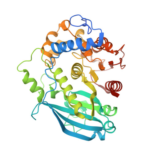
    Glucuronoyl esterases (GEs) are α/β serine hydrolases and a relatively new addition in the toolbox to reduce the recalcitrance of lignocellulose, the biggest obstacle in cost-effective utilization of this important renewable resource. While biochemical and structural characterization of GEs have progressed greatly recently, there have yet been no mechanistic studies shedding light onto the rate-limiting steps relevant for biomass conversion. The bacterial GE OtCE15A possesses a classical yet distinctive catalytic machinery, with easily identifiable catalytic Ser/His completed by two acidic residues (Glu and Asp) rather than one as in the classical triad, and an Arg side chain participating in the oxyanion hole. By QM/MM calculations, we identified deacylation as the decisive step in catalysis, and quantified the role of Asp, Glu and Arg, showing the latter to be particularly important. The results agree well with experimental and structural data. We further calculated the free-energy barrier of post-catalysis dissociation from a complex natural substrate, suggesting that in industrial settings non-catalytic processes may constitute the rate-limiting step, and pointing to future directions for enzyme engineering in biomass utilization.


  • Organizational Affiliation

    Department of Chemistry, University of Copenhagen, DK-2100, Copenhagen, Denmark.


Macromolecules
Find similar proteins by:  (by identity cutoff)  |  3D Structure
Entity ID: 1
MoleculeChains Sequence LengthOrganismDetailsImage
Putative acetyl xylan esterase421Opitutus terrae PB90-1Mutation(s): 1 
Gene Names: Oter_0116
UniProt
Find proteins for B1ZMF4 (Opitutus terrae (strain DSM 11246 / JCM 15787 / PB90-1))
Explore B1ZMF4 
Go to UniProtKB:  B1ZMF4
Entity Groups  
Sequence Clusters30% Identity50% Identity70% Identity90% Identity95% Identity100% Identity
UniProt GroupB1ZMF4
Sequence Annotations
Expand
  • Reference Sequence
Small Molecules
Ligands 5 Unique
IDChains Name / Formula / InChI Key2D Diagram3D Interactions
GCU (Subject of Investigation/LOI)
Query on GCU

Download Ideal Coordinates CCD File 
B [auth A]alpha-D-glucopyranuronic acid
C6 H10 O7
AEMOLEFTQBMNLQ-WAXACMCWSA-N
DMS
Query on DMS

Download Ideal Coordinates CCD File 
C [auth A]
D [auth A]
E [auth A]
F [auth A]
G [auth A]
C [auth A],
D [auth A],
E [auth A],
F [auth A],
G [auth A],
H [auth A],
I [auth A],
J [auth A],
K [auth A],
L [auth A],
M [auth A]
DIMETHYL SULFOXIDE
C2 H6 O S
IAZDPXIOMUYVGZ-UHFFFAOYSA-N
GLY
Query on GLY

Download Ideal Coordinates CCD File 
BA [auth A],
CA [auth A],
DA [auth A],
EA [auth A]
GLYCINE
C2 H5 N O2
DHMQDGOQFOQNFH-UHFFFAOYSA-N
EDO
Query on EDO

Download Ideal Coordinates CCD File 
AA [auth A]
P [auth A]
Q [auth A]
R [auth A]
S [auth A]
AA [auth A],
P [auth A],
Q [auth A],
R [auth A],
S [auth A],
T [auth A],
U [auth A],
V [auth A],
W [auth A],
X [auth A],
Y [auth A],
Z [auth A]
1,2-ETHANEDIOL
C2 H6 O2
LYCAIKOWRPUZTN-UHFFFAOYSA-N
NA
Query on NA

Download Ideal Coordinates CCD File 
N [auth A],
O [auth A]
SODIUM ION
Na
FKNQFGJONOIPTF-UHFFFAOYSA-N
Experimental Data & Validation

Experimental Data

  • Method: X-RAY DIFFRACTION
  • Resolution: 1.80 Å
  • R-Value Free: 0.222 
  • R-Value Work: 0.183 
  • R-Value Observed: 0.185 
  • Space Group: P 1
Unit Cell:
Length ( Å )Angle ( ˚ )
a = 47.333α = 61.947
b = 47.298β = 67.92
c = 51.112γ = 88.272
Software Package:
Software NamePurpose
PHENIXrefinement
XDSdata reduction
PHASERphasing
XSCALEdata scaling

Structure Validation

View Full Validation Report


Ligand Structure Quality Assessment 


Entry History & Funding Information

Deposition Data


Funding OrganizationLocationGrant Number
Novo Nordisk FoundationDenmarkNNF17OC0027698
Knut and Alice Wallenberg FoundationSweden--

Revision History  (Full details and data files)

  • Version 1.0: 2022-01-12
    Type: Initial release
  • Version 1.1: 2022-04-06
    Changes: Database references
  • Version 1.2: 2024-01-31
    Changes: Data collection, Refinement description