Mutation-induced dimerization of transforming growth factor-beta-induced protein may drive protein aggregation in granular corneal dystrophy.
Nielsen, N.S., Gadeberg, T.A.F., Poulsen, E.T., Harwood, S.L., Weberskov, C.E., Pedersen, J.S., Andersen, G.R., Enghild, J.J.(2021) J Biol Chem 297: 100858-100858
- PubMed: 34097874
- DOI: https://doi.org/10.1016/j.jbc.2021.100858
- Primary Citation of Related Structures:
7AS7, 7ASC, 7ASG - PubMed Abstract:
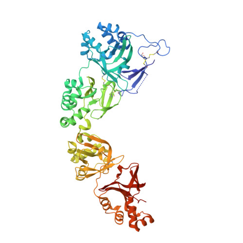
Protein aggregation in the outermost layers of the cornea, which can lead to cloudy vision and in severe cases blindness, is linked to mutations in the extracellular matrix protein transforming growth factor-β-induced protein (TGFBIp). Among the most frequent pathogenic mutations are R124H and R555W, both associated with granular corneal dystrophy (GCD) characterized by the early-onset formation of amorphous aggregates. The molecular mechanisms of protein aggregation in GCD are largely unknown. In this study, we determined the crystal structures of R124H, R555W, and the lattice corneal dystrophy-associated A546T. Although there were no changes in the monomeric TGFBIp structure of any mutant that would explain their propensity to aggregate, R124H and R555W demonstrated a new dimer interface in the crystal packing, which is not present in wildtype TGFBIp or A546T. This interface, as seen in both the R124H and R555W structures, involves residue 124 of the first TGFBIp molecule and 555 in the second. The interface is not permitted by the Arg124 and Arg555 residues of wildtype TGFBIp and may play a central role in the aggregation exhibited by R124H and R555W in vivo. Using cross-linking mass spectrometry and in-line size exclusion chromatography-small-angle X-ray scattering, we characterized a dimer formed by wildtype and mutant TGFBIps in solution. Dimerization in solution also involves interactions between the N- and C-terminal domains of two TGFBIp molecules but was not identical to the crystal packing dimerization. TGFBIp-targeted interventions that disrupt the R124H/R555W crystal packing dimer interface might offer new therapeutic opportunities to treat patients with GCD.
Organizational Affiliation:
Department of Molecular Biology and Genetics, Science Park, Aarhus University, Aarhus, Denmark.