7AES

Human carbonic anhydrase II in complex with 2,3,5,6-tetrafluoro-N-methyl-4-propylsulfanyl-benzenesulfonamide


Experimental Data Snapshot

  • Method: X-RAY DIFFRACTION
  • Resolution: 1.40 Å
  • R-Value Free: 0.209 
  • R-Value Work: 0.170 
  • R-Value Observed: 0.174 

wwPDB Validation   3D Report Full Report


This is version 1.2 of the entry. See complete history


Literature

Structure and mechanism of secondary sulfonamide binding to carbonic anhydrases.

Baronas, D.Dudutiene, V.Paketuryte, V.Kairys, V.Smirnov, A.Juozapaitiene, V.Vaskevicius, A.Manakova, E.Grazulis, S.Zubriene, A.Matulis, D.

(2021) Eur Biophys J 50: 993-1011

  • DOI: https://doi.org/10.1007/s00249-021-01561-1
  • Primary Citation of Related Structures:  
    7AEQ, 7AES, 7AGN

  • PubMed Abstract: 

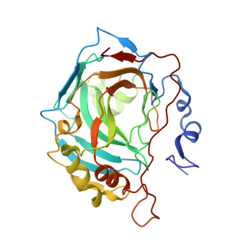
    Zinc-containing metalloenzyme carbonic anhydrase (CA) binds primary sulfonamides with extremely high, up to picomolar, affinity by forming a coordination bond between the negatively charged amino group and the zinc ion and making hydrogen bonds and hydrophobic contacts with other parts of the inhibitor molecule. However, N-methyl-substituted, secondary or tertiary sulfonamides bind CA with much lower affinity. In search for an explanation for this diminished affinity, a series of secondary sulfonamides were synthesized and, together with analogous primary sulfonamides, the affinities for 12 recombinant catalytically active human CA isoforms were determined by the fluorescent thermal shift assay, stopped-flow assay of the inhibition of enzymatic activity and isothermal titration calorimetry. The binding profile of secondary sulfonamides as a function of pH showed the same U-shape dependence seen for primary sulfonamides. This dependence demonstrated that there were protein binding-linked protonation reactions that should be dissected for the estimation of the intrinsic binding constants to perform structure-thermodynamics analysis. X-ray crystallographic structures of secondary sulfonamides and computational modeling dissected the atomic contributions to the binding energetics. Secondary sulfonamides bind to carbonic anhydrases via coordination bond between the negatively charged nitrogen of alkylated amino group and Zn(II) in the active site of CA. The binding reaction is linked to deprotonation of the amino group and protonation of the Zn(II)-bound hydroxide. To perform the structure-thermodynamics analysis, contributions of these linked reactions must be subtracted to determine the intrinsic energetics. In this aspect, the secondary sulfonamides are similar to primary sulfonamides as CA inhibitors.


  • Organizational Affiliation

    Department of Biothermodynamics and Drug Design, Institute of Biotechnology, Life Sciences Center, Vilnius University, Saulėtekio 7, 10257, Vilnius, Lithuania.


Macromolecules
Find similar proteins by:  (by identity cutoff)  |  3D Structure
Entity ID: 1
MoleculeChains Sequence LengthOrganismDetailsImage
carbonic anhydrase 2260Homo sapiensMutation(s): 0 
EC: 4.2.1.1
UniProt & NIH Common Fund Data Resources
Find proteins for P00918 (Homo sapiens)
Explore P00918 
Go to UniProtKB:  P00918
PHAROS:  P00918
GTEx:  ENSG00000104267 
Entity Groups  
Sequence Clusters30% Identity50% Identity70% Identity90% Identity95% Identity100% Identity
UniProt GroupP00918
Sequence Annotations
Expand
  • Reference Sequence
Small Molecules
Ligands 5 Unique
IDChains Name / Formula / InChI Key2D Diagram3D Interactions
R8Q (Subject of Investigation/LOI)
Query on R8Q

Download Ideal Coordinates CCD File 
C [auth A]2,3,5,6-tetrakis(fluoranyl)-~{N}-methyl-4-propylsulfanyl-benzenesulfonamide
C10 H11 F4 N O2 S2
NTKHWXIITXMCEJ-UHFFFAOYSA-N
MES
Query on MES

Download Ideal Coordinates CCD File 
D [auth A]2-(N-MORPHOLINO)-ETHANESULFONIC ACID
C6 H13 N O4 S
SXGZJKUKBWWHRA-UHFFFAOYSA-N
BCN
Query on BCN

Download Ideal Coordinates CCD File 
E [auth A]BICINE
C6 H13 N O4
FSVCELGFZIQNCK-UHFFFAOYSA-N
ZN
Query on ZN

Download Ideal Coordinates CCD File 
B [auth A]ZINC ION
Zn
PTFCDOFLOPIGGS-UHFFFAOYSA-N
NA
Query on NA

Download Ideal Coordinates CCD File 
F [auth A],
G [auth A]
SODIUM ION
Na
FKNQFGJONOIPTF-UHFFFAOYSA-N
Experimental Data & Validation

Experimental Data

  • Method: X-RAY DIFFRACTION
  • Resolution: 1.40 Å
  • R-Value Free: 0.209 
  • R-Value Work: 0.170 
  • R-Value Observed: 0.174 
  • Space Group: P 1 21 1
Unit Cell:
Length ( Å )Angle ( ˚ )
a = 42.165α = 90
b = 40.995β = 104
c = 71.752γ = 90
Software Package:
Software NamePurpose
SCALAdata scaling
REFMACrefinement
PDB_EXTRACTdata extraction
XDSdata reduction
MOLREPphasing
Cootmodel building

Structure Validation

View Full Validation Report


Entry History 

Deposition Data

Revision History  (Full details and data files)

  • Version 1.0: 2021-09-01
    Type: Initial release
  • Version 1.1: 2021-09-29
    Changes: Data collection, Database references
  • Version 1.2: 2024-01-31
    Changes: Data collection, Refinement description