6YTP

Structure of recombinant human beta-glucocerebrosidase in complex with azide tagged cyclophellitol epoxide inhibitor


Experimental Data Snapshot

  • Method: X-RAY DIFFRACTION
  • Resolution: 1.70 Å
  • R-Value Free: 0.212 
  • R-Value Work: 0.186 

wwPDB Validation   3D Report Full Report


Ligand Structure Quality Assessment 


This is version 1.3 of the entry. See complete history


Literature

Design, Synthesis and Structural Analysis of Glucocerebrosidase Imaging Agents.

Rowland, R.J.Chen, Y.Breen, I.Wu, L.Offen, W.A.Beenakker, T.J.Su, Q.van den Nieuwendijk, A.M.C.H.Aerts, J.M.F.G.Artola, M.Overkleeft, H.S.Davies, G.J.

(2021) Chemistry 27: 16377-16388

  • DOI: https://doi.org/10.1002/chem.202102359
  • Primary Citation of Related Structures:  
    6YTP, 6YTR, 6YUT, 6YV3, 6Z39, 6Z3I

  • PubMed Abstract: 

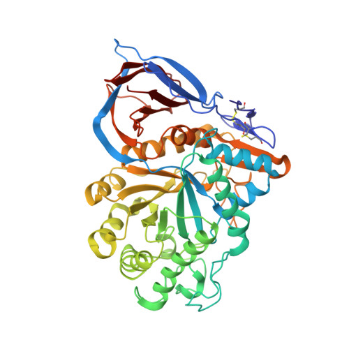
    Gaucher disease (GD) is a lysosomal storage disorder caused by inherited deficiencies in β-glucocerebrosidase (GBA). Current treatments require rapid disease diagnosis and a means of monitoring therapeutic efficacy, both of which may be supported by the use of GBA-targeting activity-based probes (ABPs). Here, we report the synthesis and structural analysis of a range of cyclophellitol epoxide and aziridine inhibitors and ABPs for GBA. We demonstrate their covalent mechanism-based mode of action and uncover binding of the new N-functionalised aziridines to the ligand binding cleft. These inhibitors became scaffolds for the development of ABPs; the O6-fluorescent tags of which bind in an allosteric site at the dimer interface. Considering GBA's preference for O6- and N-functionalised reagents, a bi-functional aziridine ABP was synthesized as a potentially more powerful imaging agent. Whilst this ABP binds to two unique active site clefts of GBA, no further benefit in potency was achieved over our first generation ABPs. Nevertheless, such ABPs should serve useful in the study of GBA in relation to GD and inform the design of future probes.


  • Organizational Affiliation

    Department of Chemistry, York Structural Biology Laboratory (YSBL), University of York Heslington, York, YO10 5DD, UK.


Macromolecules
Find similar proteins by:  (by identity cutoff)  |  3D Structure
Entity ID: 1
MoleculeChains Sequence LengthOrganismDetailsImage
GlucosylceramidaseA [auth AAA],
B [auth BBB]
497Homo sapiensMutation(s): 0 
Gene Names: GBAGCGLUC
EC: 3.2.1.45 (PDB Primary Data), 2.4.1 (PDB Primary Data), 3.2.1.104 (PDB Primary Data)
UniProt & NIH Common Fund Data Resources
Find proteins for P04062 (Homo sapiens)
Explore P04062 
Go to UniProtKB:  P04062
PHAROS:  P04062
GTEx:  ENSG00000177628 
Entity Groups  
Sequence Clusters30% Identity50% Identity70% Identity90% Identity95% Identity100% Identity
UniProt GroupP04062
Sequence Annotations
Expand
  • Reference Sequence
Oligosaccharides

Help

Entity ID: 2
MoleculeChains Length2D Diagram Glycosylation3D Interactions
2-acetamido-2-deoxy-beta-D-glucopyranose-(1-4)-2-acetamido-2-deoxy-beta-D-glucopyranoseC [auth A],
D [auth B]
2N-Glycosylation
Glycosylation Resources
GlyTouCan:  G42666HT
GlyCosmos:  G42666HT
GlyGen:  G42666HT
Small Molecules
Ligands 5 Unique
IDChains Name / Formula / InChI Key2D Diagram3D Interactions
NAG
Query on NAG

Download Ideal Coordinates CCD File 
E [auth AAA],
X [auth BBB]
2-acetamido-2-deoxy-beta-D-glucopyranose
C8 H15 N O6
OVRNDRQMDRJTHS-FMDGEEDCSA-N
PN8 (Subject of Investigation/LOI)
Query on PN8

Download Ideal Coordinates CCD File 
PA [auth BBB],
W [auth AAA]
(1~{S},2~{R},3~{R},4~{S},5~{S})-4-[[($l^{5}-azanylidyne-$l^{5}-azanyl)amino]methyl]cyclohexane-1,2,3,5-tetrol
C7 H14 N3 O4
FBFTWYYGVGMNEW-YPPKRKOXSA-N
SO4
Query on SO4

Download Ideal Coordinates CCD File 
AA [auth BBB]
BA [auth BBB]
CA [auth BBB]
DA [auth BBB]
EA [auth BBB]
AA [auth BBB],
BA [auth BBB],
CA [auth BBB],
DA [auth BBB],
EA [auth BBB],
F [auth AAA],
FA [auth BBB],
G [auth AAA],
H [auth AAA],
I [auth AAA],
J [auth AAA],
K [auth AAA],
L [auth AAA],
Y [auth BBB],
Z [auth BBB]
SULFATE ION
O4 S
QAOWNCQODCNURD-UHFFFAOYSA-L
GOL
Query on GOL

Download Ideal Coordinates CCD File 
KA [auth BBB]
LA [auth BBB]
MA [auth BBB]
NA [auth BBB]
OA [auth BBB]
KA [auth BBB],
LA [auth BBB],
MA [auth BBB],
NA [auth BBB],
OA [auth BBB],
U [auth AAA],
V [auth AAA]
GLYCEROL
C3 H8 O3
PEDCQBHIVMGVHV-UHFFFAOYSA-N
EDO
Query on EDO

Download Ideal Coordinates CCD File 
GA [auth BBB]
HA [auth BBB]
IA [auth BBB]
JA [auth BBB]
M [auth AAA]
GA [auth BBB],
HA [auth BBB],
IA [auth BBB],
JA [auth BBB],
M [auth AAA],
N [auth AAA],
O [auth AAA],
P [auth AAA],
Q [auth AAA],
R [auth AAA],
S [auth AAA],
T [auth AAA]
1,2-ETHANEDIOL
C2 H6 O2
LYCAIKOWRPUZTN-UHFFFAOYSA-N
Experimental Data & Validation

Experimental Data

  • Method: X-RAY DIFFRACTION
  • Resolution: 1.70 Å
  • R-Value Free: 0.212 
  • R-Value Work: 0.186 
  • Space Group: C 2 2 21
Unit Cell:
Length ( Å )Angle ( ˚ )
a = 108.89α = 90
b = 285.769β = 90
c = 91.674γ = 90
Software Package:
Software NamePurpose
REFMACrefinement
xia2data reduction
Aimlessdata scaling
MOLREPphasing

Structure Validation

View Full Validation Report


Ligand Structure Quality Assessment 


Entry History & Funding Information

Deposition Data


Funding OrganizationLocationGrant Number
Biotechnology and Biological Sciences Research Council (BBSRC)United KingdomBB/M011151/1

Revision History  (Full details and data files)

  • Version 1.0: 2021-05-12
    Type: Initial release
  • Version 1.1: 2021-11-24
    Changes: Data collection, Database references, Structure summary
  • Version 1.2: 2021-12-08
    Changes: Data collection, Database references
  • Version 1.3: 2024-01-24
    Changes: Data collection, Derived calculations, Refinement description