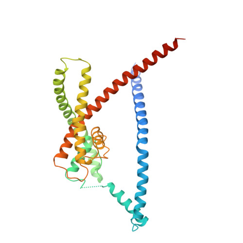
6W88

K2P2.1 (TREK-1):ML335 complex, 30 mM K+


Experimental Data Snapshot

  • Method: X-RAY DIFFRACTION
  • Resolution: 3.20 Å
  • R-Value Free: 0.294 
  • R-Value Work: 0.259 
  • R-Value Observed: 0.261 

wwPDB Validation   3D Report Full Report


Ligand Structure Quality Assessment 


This is version 1.1 of the entry. See complete history


Literature

K 2P channel C-type gating involves asymmetric selectivity filter order-disorder transitions.

Lolicato, M.Natale, A.M.Abderemane-Ali, F.Crottes, D.Capponi, S.Duman, R.Wagner, A.Rosenberg, J.M.Grabe, M.Minor Jr., D.L.

(2020) Sci Adv 6

  • DOI: https://doi.org/10.1126/sciadv.abc9174
  • Primary Citation of Related Structures:  
    6W7D, 6W7E, 6W83, 6W84, 6W85, 6W86, 6W87, 6W88, 6W8A, 6W8F

  • PubMed Abstract: 

    K 2P potassium channels regulate cellular excitability using their selectivity filter (C-type) gate. C-type gating mechanisms, best characterized in homotetrameric potassium channels, remain controversial and are attributed to selectivity filter pinching, dilation, or subtle structural changes. The extent to which such mechanisms control C-type gating of innately heterodimeric K 2P s is unknown. Here, combining K 2P 2.1 (TREK-1) x-ray crystallography in different potassium concentrations, potassium anomalous scattering, molecular dynamics, and electrophysiology, we uncover unprecedented, asymmetric, potassium-dependent conformational changes that underlie K 2P C-type gating. These asymmetric order-disorder transitions, enabled by the K 2P heterodimeric architecture, encompass pinching and dilation, disrupt the S1 and S2 ion binding sites, require the uniquely long K 2P SF2-M4 loop and conserved "M3 glutamate network," and are suppressed by the K 2P C-type gate activator ML335. These findings demonstrate that two distinct C-type gating mechanisms can operate in one channel and underscore the SF2-M4 loop as a target for K 2P channel modulator development.


  • Organizational Affiliation

    Cardiovascular Research Institute, University of California, San Francisco, CA 93858-2330, USA.


Macromolecules
Find similar proteins by:  (by identity cutoff)  |  3D Structure
Entity ID: 1
MoleculeChains Sequence LengthOrganismDetailsImage
Potassium channel subfamily K member 2
A, B
312Mus musculusMutation(s): 13 
Gene Names: Kcnk2
Membrane Entity: Yes 
UniProt & NIH Common Fund Data Resources
Find proteins for P97438 (Mus musculus)
Explore P97438 
Go to UniProtKB:  P97438
IMPC:  MGI:109366
Entity Groups  
Sequence Clusters30% Identity50% Identity70% Identity90% Identity95% Identity100% Identity
UniProt GroupP97438
Sequence Annotations
Expand
  • Reference Sequence
Small Molecules
Ligands 9 Unique
IDChains Name / Formula / InChI Key2D Diagram3D Interactions
Q6F
Query on Q6F

Download Ideal Coordinates CCD File 
C [auth A],
T [auth B]
N-[(2,4-dichlorophenyl)methyl]-4-[(methylsulfonyl)amino]benzamide
C15 H14 Cl2 N2 O3 S
RDFIQTZRJRVFHK-UHFFFAOYSA-N
R16
Query on R16

Download Ideal Coordinates CCD File 
AA [auth B],
J [auth A],
Z [auth B]
HEXADECANE
C16 H34
DCAYPVUWAIABOU-UHFFFAOYSA-N
D12
Query on D12

Download Ideal Coordinates CCD File 
E [auth A],
F [auth A],
U [auth B]
DODECANE
C12 H26
SNRUBQQJIBEYMU-UHFFFAOYSA-N
UND
Query on UND

Download Ideal Coordinates CCD File 
O [auth A],
P [auth A]
UNDECANE
C11 H24
RSJKGSCJYJTIGS-UHFFFAOYSA-N
D10
Query on D10

Download Ideal Coordinates CCD File 
G [auth A],
V [auth B]
DECANE
C10 H22
DIOQZVSQGTUSAI-UHFFFAOYSA-N
OCT
Query on OCT

Download Ideal Coordinates CCD File 
I [auth A],
X [auth B],
Y [auth B]
N-OCTANE
C8 H18
TVMXDCGIABBOFY-UHFFFAOYSA-N
CD
Query on CD

Download Ideal Coordinates CCD File 
D [auth A],
Q [auth B],
R [auth B],
S [auth B]
CADMIUM ION
Cd
WLZRMCYVCSSEQC-UHFFFAOYSA-N
LNK
Query on LNK

Download Ideal Coordinates CCD File 
H [auth A],
W [auth B]
PENTANE
C5 H12
OFBQJSOFQDEBGM-UHFFFAOYSA-N
K
Query on K

Download Ideal Coordinates CCD File 
BA [auth B],
K [auth A],
L [auth A],
M [auth A],
N [auth A]
POTASSIUM ION
K
NPYPAHLBTDXSSS-UHFFFAOYSA-N
Experimental Data & Validation

Experimental Data

  • Method: X-RAY DIFFRACTION
  • Resolution: 3.20 Å
  • R-Value Free: 0.294 
  • R-Value Work: 0.259 
  • R-Value Observed: 0.261 
  • Space Group: P 21 21 21
Unit Cell:
Length ( Å )Angle ( ˚ )
a = 67.006α = 90
b = 119.996β = 90
c = 128.794γ = 90
Software Package:
Software NamePurpose
BUSTERrefinement
XDSdata reduction
Aimlessdata scaling
PHASERphasing

Structure Validation

View Full Validation Report


Ligand Structure Quality Assessment 


Entry History & Funding Information

Deposition Data


Funding OrganizationLocationGrant Number
National Institutes of Health/National Institute of Mental Health (NIH/NIMH)United StatesNIH-R01-MH093603

Revision History  (Full details and data files)

  • Version 1.0: 2021-01-27
    Type: Initial release
  • Version 1.1: 2023-10-18
    Changes: Data collection, Database references, Refinement description