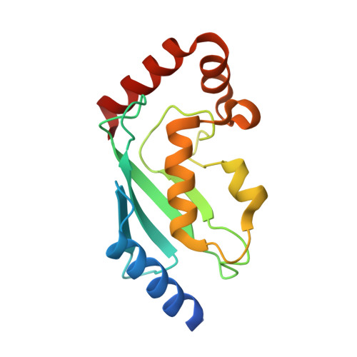
Dimerization regulates the human APC/C-associated ubiquitin-conjugating enzyme UBE2S.
Liess, A.K.L., Kucerova, A., Schweimer, K., Schlesinger, D., Dybkov, O., Urlaub, H., Mansfeld, J., Lorenz, S.(2020) Sci Signal 13
- PubMed: 33082289
- DOI: https://doi.org/10.1126/scisignal.aba8208
- Primary Citation of Related Structures:
6S96, 6S98 - PubMed Abstract:
At the heart of protein ubiquitination cascades, ubiquitin-conjugating enzymes (E2s) form reactive ubiquitin-thioester intermediates to enable efficient transfer of ubiquitin to cellular substrates. The precise regulation of E2s is thus crucial for cellular homeostasis, and their deregulation is frequently associated with tumorigenesis. In addition to driving substrate ubiquitination together with ubiquitin ligases (E3s), many E2s can also autoubiquitinate, thereby promoting their own proteasomal turnover. To investigate the mechanisms that balance these disparate activities, we dissected the regulatory dynamics of UBE2S, a human APC/C-associated E2 that ensures the faithful ubiquitination of cell cycle regulators during mitosis. We uncovered a dimeric state of UBE2S that confers autoinhibition by blocking a catalytically critical ubiquitin binding site. Dimerization is stimulated by the lysine-rich carboxyl-terminal extension of UBE2S that is also required for the recruitment of this E2 to the APC/C and is autoubiquitinated as substrate abundance becomes limiting. Consistent with this mechanism, we found that dimerization-deficient UBE2S turned over more rapidly in cells and did not promote mitotic slippage during prolonged drug-induced mitotic arrest. We propose that dimerization attenuates the autoubiquitination-induced turnover of UBE2S when the APC/C is not fully active. More broadly, our data illustrate how the use of mutually exclusive macromolecular interfaces enables modulation of both the activities and the abundance of E2s in cells to facilitate precise ubiquitin signaling.
Organizational Affiliation:
Rudolf Virchow Center for Integrative and Translational Bioimaging, University of Würzburg, 97080 Würzburg, Germany.