6RV6

Structure of properdin lacking TSR3 based on anomalous data


Experimental Data Snapshot

  • Method: X-RAY DIFFRACTION
  • Resolution: 3.51 Å
  • R-Value Free: 0.283 
  • R-Value Work: 0.264 
  • R-Value Observed: 0.265 

wwPDB Validation   3D Report Full Report


Ligand Structure Quality Assessment 


This is version 2.0 of the entry. See complete history


Literature

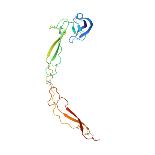
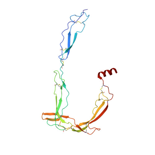
Structural Basis for Properdin Oligomerization and Convertase Stimulation in the Human Complement System.

Pedersen, D.V.Gadeberg, T.A.F.Thomas, C.Wang, Y.Joram, N.Jensen, R.K.Mazarakis, S.M.M.Revel, M.El Sissy, C.Petersen, S.V.Lindorff-Larsen, K.Thiel, S.Laursen, N.S.Fremeaux-Bacchi, V.Andersen, G.R.

(2019) Front Immunol 10: 2007-2007

  • DOI: https://doi.org/10.3389/fimmu.2019.02007
  • Primary Citation of Related Structures:  
    6RU3, 6RU5, 6RUR, 6RUS, 6RUV, 6RV6, 6SEJ

  • PubMed Abstract: 

    Properdin (FP) is a positive regulator of the immune system stimulating the activity of the proteolytically active C3 convertase C3bBb in the alternative pathway of the complement system. Here we present two crystal structures of FP and two structures of convertase bound FP. A structural core formed by three thrombospondin repeats (TSRs) and a TB domain harbors the convertase binding site in FP that mainly interacts with C3b. Stabilization of the interaction between the C3b C-terminus and the MIDAS bound Mg 2+ in the Bb protease by FP TSR5 is proposed to underlie FP convertase stabilization. Intermolecular contacts between FP and the convertase subunits suggested by the structure were confirmed by binding experiments. FP is shown to inhibit C3b degradation by FI due to a direct competition for a common binding site on C3b. FP oligomers are held together by two sets of intermolecular contacts, where the first is formed by the TB domain from one FP molecule and TSR4 from another. The second and largest interface is formed by TSR1 and TSR6 from the same two FP molecules. Flexibility at four hinges between thrombospondin repeats is suggested to enable the oligomeric, polydisperse, and extended architecture of FP. Our structures rationalize the effects of mutations associated with FP deficiencies and provide a structural basis for the analysis of FP function in convertases and its possible role in pattern recognition.


  • Organizational Affiliation

    Department of Molecular Biology and Genetics, Center for Structural Biology, Aarhus University, Aarhus, Denmark.


Macromolecules
Find similar proteins by:  (by identity cutoff)  |  3D Structure
Entity ID: 1
MoleculeChains Sequence LengthOrganismDetailsImage
Properdin170Homo sapiensMutation(s): 0 
Gene Names: CFPPFC
UniProt & NIH Common Fund Data Resources
Find proteins for P27918 (Homo sapiens)
Explore P27918 
Go to UniProtKB:  P27918
PHAROS:  P27918
GTEx:  ENSG00000126759 
Entity Groups  
Sequence Clusters30% Identity50% Identity70% Identity90% Identity95% Identity100% Identity
UniProt GroupP27918
Sequence Annotations
Expand
  • Reference Sequence
Find similar proteins by:  (by identity cutoff)  |  3D Structure
Entity ID: 2
MoleculeChains Sequence LengthOrganismDetailsImage
Properdin221Homo sapiensMutation(s): 0 
Gene Names: CFPPFC
UniProt & NIH Common Fund Data Resources
Find proteins for P27918 (Homo sapiens)
Explore P27918 
Go to UniProtKB:  P27918
PHAROS:  P27918
GTEx:  ENSG00000126759 
Entity Groups  
Sequence Clusters30% Identity50% Identity70% Identity90% Identity95% Identity100% Identity
UniProt GroupP27918
Sequence Annotations
Expand
  • Reference Sequence
Oligosaccharides

Help

Entity ID: 3
MoleculeChains Length2D Diagram Glycosylation3D Interactions
beta-D-glucopyranose-(1-3)-alpha-L-fucopyranose
C, D, E
2O-Glycosylation
Glycosylation Resources
GlyTouCan:  G36855WW
GlyCosmos:  G36855WW
GlyGen:  G36855WW
Entity ID: 4
MoleculeChains Length2D Diagram Glycosylation3D Interactions
2-acetamido-2-deoxy-beta-D-glucopyranose-(1-4)-[alpha-L-fucopyranose-(1-6)]2-acetamido-2-deoxy-beta-D-glucopyranose
F
3N-Glycosylation
Glycosylation Resources
GlyTouCan:  G21290RB
GlyCosmos:  G21290RB
GlyGen:  G21290RB
Small Molecules
Ligands 1 Unique
IDChains Name / Formula / InChI Key2D Diagram3D Interactions
MAN
Query on MAN

Download Ideal Coordinates CCD File 
G [auth A]
H [auth A]
I [auth A]
J [auth A]
K [auth A]
G [auth A],
H [auth A],
I [auth A],
J [auth A],
K [auth A],
L [auth B],
M [auth B],
N [auth B],
O [auth B],
P [auth B],
Q [auth B],
R [auth B]
alpha-D-mannopyranose
C6 H12 O6
WQZGKKKJIJFFOK-PQMKYFCFSA-N
Experimental Data & Validation

Experimental Data

  • Method: X-RAY DIFFRACTION
  • Resolution: 3.51 Å
  • R-Value Free: 0.283 
  • R-Value Work: 0.264 
  • R-Value Observed: 0.265 
  • Space Group: I 41
Unit Cell:
Length ( Å )Angle ( ˚ )
a = 219.79α = 90
b = 219.79β = 90
c = 47.44γ = 90
Software Package:
Software NamePurpose
PHENIXrefinement
XDSdata reduction
XSCALEdata scaling
PHASERphasing

Structure Validation

View Full Validation Report


Ligand Structure Quality Assessment 


Entry History & Funding Information

Deposition Data


Funding OrganizationLocationGrant Number
LundbeckfondenDenmarkR155-2015-2666

Revision History  (Full details and data files)

  • Version 1.0: 2019-08-21
    Type: Initial release
  • Version 1.1: 2019-09-25
    Changes: Data collection, Database references
  • Version 2.0: 2020-07-29
    Type: Remediation
    Reason: Carbohydrate remediation
    Changes: Advisory, Atomic model, Data collection, Derived calculations, Structure summary