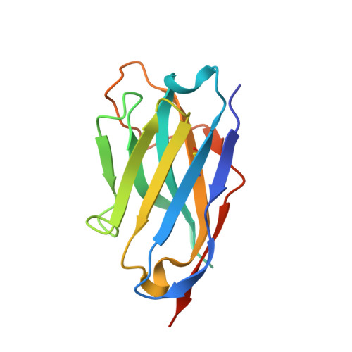
Structural Basis for Properdin Oligomerization and Convertase Stimulation in the Human Complement System.
Pedersen, D.V., Gadeberg, T.A.F., Thomas, C., Wang, Y., Joram, N., Jensen, R.K., Mazarakis, S.M.M., Revel, M., El Sissy, C., Petersen, S.V., Lindorff-Larsen, K., Thiel, S., Laursen, N.S., Fremeaux-Bacchi, V., Andersen, G.R.(2019) Front Immunol 10: 2007-2007
- PubMed: 31507604
- DOI: https://doi.org/10.3389/fimmu.2019.02007
- Primary Citation of Related Structures:
6RU3, 6RU5, 6RUR, 6RUS, 6RUV, 6RV6, 6SEJ - PubMed Abstract:
Properdin (FP) is a positive regulator of the immune system stimulating the activity of the proteolytically active C3 convertase C3bBb in the alternative pathway of the complement system. Here we present two crystal structures of FP and two structures of convertase bound FP. A structural core formed by three thrombospondin repeats (TSRs) and a TB domain harbors the convertase binding site in FP that mainly interacts with C3b. Stabilization of the interaction between the C3b C-terminus and the MIDAS bound Mg 2+ in the Bb protease by FP TSR5 is proposed to underlie FP convertase stabilization. Intermolecular contacts between FP and the convertase subunits suggested by the structure were confirmed by binding experiments. FP is shown to inhibit C3b degradation by FI due to a direct competition for a common binding site on C3b. FP oligomers are held together by two sets of intermolecular contacts, where the first is formed by the TB domain from one FP molecule and TSR4 from another. The second and largest interface is formed by TSR1 and TSR6 from the same two FP molecules. Flexibility at four hinges between thrombospondin repeats is suggested to enable the oligomeric, polydisperse, and extended architecture of FP. Our structures rationalize the effects of mutations associated with FP deficiencies and provide a structural basis for the analysis of FP function in convertases and its possible role in pattern recognition.
Organizational Affiliation:
Department of Molecular Biology and Genetics, Center for Structural Biology, Aarhus University, Aarhus, Denmark.


