6R1I

Structure of porcine Aichi virus polymerase


Experimental Data Snapshot

  • Method: X-RAY DIFFRACTION
  • Resolution: 2.63 Å
  • R-Value Free: 0.222 
  • R-Value Work: 0.191 
  • R-Value Observed: 0.192 

wwPDB Validation   3D Report Full Report


This is version 1.2 of the entry. See complete history


Literature

Structures of kobuviral and siciniviral polymerases reveal conserved mechanism of picornaviral polymerase activation.

Dubankova, A.Horova, V.Klima, M.Boura, E.

(2019) J Struct Biol 208: 92-98

  • DOI: https://doi.org/10.1016/j.jsb.2019.08.004
  • Primary Citation of Related Structures:  
    6QWT, 6R1I

  • PubMed Abstract: 

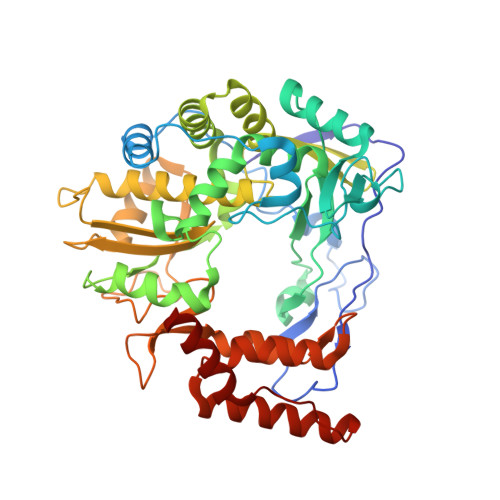
    RNA-dependent RNA polymerase 3D pol is a key enzyme for the replication of picornaviruses. The viral genome is translated into a single polyprotein that is subsequently proteolytically processed into matured products. The 3D pol enzyme arises from a stable 3CD precursor that has high proteolytic activity but no polymerase activity. Upon cleavage of the precursor the newly established N-terminus of 3D pol is liberated and inserts itself into a pocket on the surface of the 3D pol enzyme. The essential residue for this mechanism is the very first glycine that is conserved among almost all picornaviruses. However, kobuviruses and siciniviruses have a serine residue instead. Intrigued by this anomaly we sought to solve the crystal structure of these 3D pol enzymes. The structures revealed a unique fold of the 3D pol N-termini but the very first serine residues were inserted into a charged pocket in a similar manner as the glycine residue in other picornaviruses. These structures revealed a common underlying mechanism of 3D pol activation that lies in activation of the α10 helix containing a key catalytical residue Asp238 that forms a hydrogen bond with the 2' hydroxyl group of the incoming NTP nucleotide.


  • Organizational Affiliation

    Institute of Organic Chemistry and Biochemistry of the Czech Academy of Sciences, Flemingovo nam. 2., 166 10 Prague 6, Czech Republic.


Macromolecules
Find similar proteins by:  (by identity cutoff)  |  3D Structure
Entity ID: 1
MoleculeChains Sequence LengthOrganismDetailsImage
Genome polyprotein
A, B
468Porcine kobuvirus swine/S-1-HUN/2007/HungaryMutation(s): 0 
UniProt
Find proteins for B8R1T8 (Porcine kobuvirus swine/S-1-HUN/2007/Hungary)
Explore B8R1T8 
Go to UniProtKB:  B8R1T8
Entity Groups  
Sequence Clusters30% Identity50% Identity70% Identity90% Identity95% Identity100% Identity
UniProt GroupB8R1T8
Sequence Annotations
Expand
  • Reference Sequence
Experimental Data & Validation

Experimental Data

  • Method: X-RAY DIFFRACTION
  • Resolution: 2.63 Å
  • R-Value Free: 0.222 
  • R-Value Work: 0.191 
  • R-Value Observed: 0.192 
  • Space Group: P 61 2 2
Unit Cell:
Length ( Å )Angle ( ˚ )
a = 170.448α = 90
b = 170.448β = 90
c = 231.339γ = 120
Software Package:
Software NamePurpose
PHENIXrefinement
XDSdata reduction
XDSdata scaling
PHASERphasing

Structure Validation

View Full Validation Report


Entry History & Funding Information

Deposition Data


Funding OrganizationLocationGrant Number
European Regional Development FundCzech RepublicCZ.02.1.01/0.0/0.0/16_019/0000729

Revision History  (Full details and data files)

  • Version 1.0: 2019-10-09
    Type: Initial release
  • Version 1.1: 2019-11-06
    Changes: Data collection, Database references
  • Version 1.2: 2024-01-24
    Changes: Data collection, Database references, Refinement description