6Q82

Crystal structure of the biportin Pdr6 in complex with RanGTP


Experimental Data Snapshot

  • Method: X-RAY DIFFRACTION
  • Resolution: 2.99 Å
  • R-Value Free: 0.253 
  • R-Value Work: 0.218 
  • R-Value Observed: 0.219 

wwPDB Validation   3D Report Full Report


Ligand Structure Quality Assessment 


This is version 1.2 of the entry. See complete history


Literature

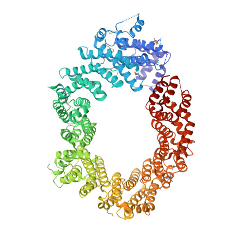
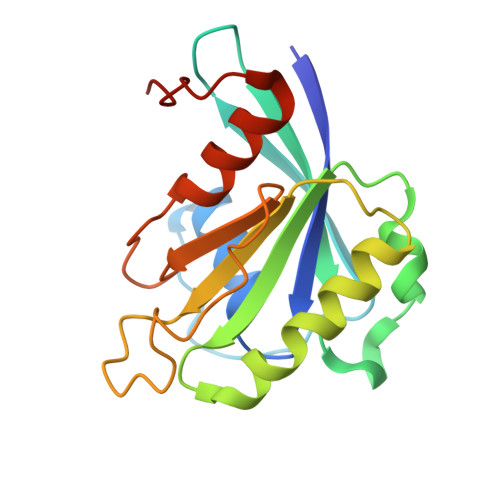
Structural basis for the nuclear import and export functions of the biportin Pdr6/Kap122.

Aksu, M.Trakhanov, S.Vera Rodriguez, A.Gorlich, D.

(2019) J Cell Biol 218: 1839-1852

  • DOI: https://doi.org/10.1083/jcb.201812093
  • Primary Citation of Related Structures:  
    6Q82, 6Q83, 6Q84

  • PubMed Abstract: 

    Importins ferry proteins into nuclei while exportins carry cargoes to the cytoplasm. In the accompanying paper in this issue (Vera Rodriguez et al. 2019. J. Cell Biol. https://doi.org/10.1083/jcb.201812091), we discovered that Pdr6 is a biportin that imports, e.g., the SUMO E2 ligase Ubc9 while depleting the translation factor eIF5A from the nuclear compartment. In this paper, we report the structures of key transport intermediates, namely, of the Ubc9•Pdr6 import complex, of the RanGTP•Pdr6 heterodimer, and of the trimeric RanGTP•Pdr6•eIF5A export complex. These revealed nonlinear transport signals, chaperone-like interactions, and how the RanGTPase system drives Pdr6 to transport Ubc9 and eIF5A in opposite directions. The structures also provide unexpected insights into the evolution of transport selectivity. Specifically, they show that recognition of Ubc9 by Pdr6 differs fundamentally from that of the human Ubc9-importer Importin 13. Likewise, Pdr6 recognizes eIF5A in a nonhomologous manner compared with the mammalian eIF5A-exporter Exportin 4. This suggests that the import of Ubc9 and active nuclear exclusion of eIF5A evolved in different eukaryotic lineages more than once and independently from each other.


  • Organizational Affiliation

    Department of Cellular Logistics, Max Planck Institute for Biophysical Chemistry, Göttingen, Germany.


Macromolecules
Find similar proteins by:  (by identity cutoff)  |  3D Structure
Entity ID: 1
MoleculeChains Sequence LengthOrganismDetailsImage
Importin beta-like protein KAP1221,080Saccharomyces cerevisiae S288CMutation(s): 0 
Gene Names: KAP122PDR6YGL016W
UniProt
Find proteins for P32767 (Saccharomyces cerevisiae (strain ATCC 204508 / S288c))
Explore P32767 
Go to UniProtKB:  P32767
Entity Groups  
Sequence Clusters30% Identity50% Identity70% Identity90% Identity95% Identity100% Identity
UniProt GroupP32767
Sequence Annotations
Expand
  • Reference Sequence
Find similar proteins by:  (by identity cutoff)  |  3D Structure
Entity ID: 2
MoleculeChains Sequence LengthOrganismDetailsImage
GTP-binding nuclear protein Ran176Homo sapiensMutation(s): 1 
Gene Names: RANARA24OK/SW-cl.81
UniProt & NIH Common Fund Data Resources
Find proteins for P62826 (Homo sapiens)
Explore P62826 
Go to UniProtKB:  P62826
PHAROS:  P62826
GTEx:  ENSG00000132341 
Entity Groups  
Sequence Clusters30% Identity50% Identity70% Identity90% Identity95% Identity100% Identity
UniProt GroupP62826
Sequence Annotations
Expand
  • Reference Sequence
Small Molecules
Ligands 2 Unique
IDChains Name / Formula / InChI Key2D Diagram3D Interactions
GTP
Query on GTP

Download Ideal Coordinates CCD File 
C [auth B]GUANOSINE-5'-TRIPHOSPHATE
C10 H16 N5 O14 P3
XKMLYUALXHKNFT-UUOKFMHZSA-N
MG
Query on MG

Download Ideal Coordinates CCD File 
D [auth B]MAGNESIUM ION
Mg
JLVVSXFLKOJNIY-UHFFFAOYSA-N
Modified Residues  1 Unique
IDChains TypeFormula2D DiagramParent
MSE
Query on MSE
A
L-PEPTIDE LINKINGC5 H11 N O2 SeMET
Experimental Data & Validation

Experimental Data

  • Method: X-RAY DIFFRACTION
  • Resolution: 2.99 Å
  • R-Value Free: 0.253 
  • R-Value Work: 0.218 
  • R-Value Observed: 0.219 
  • Space Group: P 63 2 2
Unit Cell:
Length ( Å )Angle ( ˚ )
a = 192.99α = 90
b = 192.99β = 90
c = 229.17γ = 120
Software Package:
Software NamePurpose
PHENIXrefinement
RESOLVEmodel building
XDSdata reduction
XSCALEdata scaling
AutoSolphasing

Structure Validation

View Full Validation Report


Ligand Structure Quality Assessment 


Entry History 

Deposition Data

Revision History  (Full details and data files)

  • Version 1.0: 2019-05-01
    Type: Initial release
  • Version 1.1: 2019-05-08
    Changes: Data collection, Database references
  • Version 1.2: 2019-06-12
    Changes: Data collection, Database references