6M9A

Bordetella pertussis globin coupled sensor regulatory domain (BpeGReg)


Experimental Data Snapshot

  • Method: X-RAY DIFFRACTION
  • Resolution: 2.30 Å
  • R-Value Free: 0.227 
  • R-Value Work: 0.161 
  • R-Value Observed: 0.167 

wwPDB Validation   3D Report Full Report


Ligand Structure Quality Assessment 


This is version 1.3 of the entry. See complete history


Literature

Structural Insights into Oxygen-Dependent Signal Transduction within Globin Coupled Sensors.

Rivera, S.Young, P.G.Hoffer, E.D.Vansuch, G.E.Metzler, C.L.Dunham, C.M.Weinert, E.E.

(2018) Inorg Chem 57: 14386-14395

  • DOI: https://doi.org/10.1021/acs.inorgchem.8b02584
  • Primary Citation of Related Structures:  
    6M9A

  • PubMed Abstract: 

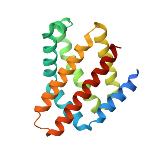
    In order to respond to external stimuli, bacteria have evolved sensor proteins linking external signals to intracellular outputs that can then regulate downstream pathways and phenotypes. Globin coupled sensor proteins (GCSs) serve to link environmental O 2 levels to cellular processes by coupling a heme-containing sensor globin domain to a catalytic output domain. However, the mechanism by which O 2 binding activates these proteins is currently unknown. To provide insights into the signaling mechanism, two distinct dimeric complexes of the isolated globin domain of the GCS from Bordetella pertussis ( BpeGlobin) were solved via X-ray crystallography in which differences in ligand-bound states were observed. Both monomers of one dimer contain Fe(II)-O 2 states, while the other dimer consists of the Fe(III)-H 2 O and Fe(II)-O 2 states. These data provide the first molecular insights into the heme pocket conformation of the active Fe(II)-O 2 form of these enzymes. In addition, heme distortion modes and heme-protein interactions were found to correlate with the ligation state of the globin, suggesting that these conformational changes play a role in O 2 -dependent signaling. Fourier transform infrared spectroscopy (FTIR) of the full-length GCS from B. pertussis ( BpeGReg) and the closely related GCS from Pectobacterium carotovorum ssp. carotovorum ( PccGCS) confirmed the importance of an ordered water within the heme pocket and two distal residues (Tyr43 and Ser68) as hydrogen-bond donors. Taken together, this work provides mechanistic insights into BpeGReg O 2 sensing and the signaling mechanisms of diguanylate cyclase-containing GCS proteins.


  • Organizational Affiliation

    Department of Chemistry , Emory University , Atlanta , Georgia 30322 , United States.


Macromolecules
Find similar proteins by:  (by identity cutoff)  |  3D Structure
Entity ID: 1
MoleculeChains Sequence LengthOrganismDetailsImage
Signaling protein
A, B, C
170Bordetella pertussisMutation(s): 0 
Gene Names: dosCERS1058649_03633
EC: 2.7.7.65
UniProt
Find proteins for Q7VTL8 (Bordetella pertussis (strain Tohama I / ATCC BAA-589 / NCTC 13251))
Explore Q7VTL8 
Go to UniProtKB:  Q7VTL8
Entity Groups  
Sequence Clusters30% Identity50% Identity70% Identity90% Identity95% Identity100% Identity
UniProt GroupQ7VTL8
Sequence Annotations
Expand
  • Reference Sequence
Small Molecules
Ligands 3 Unique
IDChains Name / Formula / InChI Key2D Diagram3D Interactions
HEM
Query on HEM

Download Ideal Coordinates CCD File 
D [auth A],
G [auth B],
J [auth C]
PROTOPORPHYRIN IX CONTAINING FE
C34 H32 Fe N4 O4
KABFMIBPWCXCRK-RGGAHWMASA-L
CA
Query on CA

Download Ideal Coordinates CCD File 
F [auth A],
I [auth B],
K [auth C]
CALCIUM ION
Ca
BHPQYMZQTOCNFJ-UHFFFAOYSA-N
PEO
Query on PEO

Download Ideal Coordinates CCD File 
E [auth A],
H [auth B]
HYDROGEN PEROXIDE
H2 O2
MHAJPDPJQMAIIY-UHFFFAOYSA-N
Experimental Data & Validation

Experimental Data

  • Method: X-RAY DIFFRACTION
  • Resolution: 2.30 Å
  • R-Value Free: 0.227 
  • R-Value Work: 0.161 
  • R-Value Observed: 0.167 
  • Space Group: C 1 2 1
Unit Cell:
Length ( Å )Angle ( ˚ )
a = 145.16α = 90
b = 49.64β = 110.58
c = 68.14γ = 90
Software Package:
Software NamePurpose
XDSdata reduction
XSCALEdata scaling
PHASERphasing
PHENIXrefinement
PDB_EXTRACTdata extraction

Structure Validation

View Full Validation Report


Ligand Structure Quality Assessment 


Entry History & Funding Information

Deposition Data


Funding OrganizationLocationGrant Number
National Science Foundation (NSF, United States)United StatesCHE1352040

Revision History  (Full details and data files)

  • Version 1.0: 2018-11-14
    Type: Initial release
  • Version 1.1: 2018-11-28
    Changes: Data collection, Database references
  • Version 1.2: 2019-11-27
    Changes: Author supporting evidence
  • Version 1.3: 2023-10-11
    Changes: Data collection, Database references, Derived calculations, Refinement description