6LJC

Crystal structure of fragmin F2-F3 domains (calcium and magnesium condition)


Experimental Data Snapshot

  • Method: X-RAY DIFFRACTION
  • Resolution: 1.85 Å
  • R-Value Free: 0.203 
  • R-Value Work: 0.182 
  • R-Value Observed: 0.183 

wwPDB Validation   3D Report Full Report


This is version 1.2 of the entry. See complete history


Literature

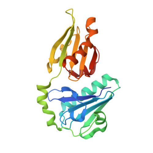
Novel inter-domain Ca2+-binding site in the gelsolin superfamily protein fragmin.

Takeda, S.Fujiwara, I.Sugimoto, Y.Oda, T.Narita, A.Maeda, Y.

(2020) J Muscle Res Cell Motil 41: 153-162

  • DOI: https://doi.org/10.1007/s10974-019-09571-5
  • Primary Citation of Related Structures:  
    6KWZ, 6LJC, 6LJD, 6LJE, 6LJF

  • PubMed Abstract: 

    Gelsolin superfamily proteins, consisting of multiple domains (usually six), sever actin filaments and cap the barbed ends in a Ca 2+ -dependent manner. Two types of evolutionally conserved Ca 2+ -binding sites have been identified in this family; type-1 (between gelsolin and actin) and type-2 (within the gelsolin domain). Fragmin, a member in the slime mold Physarum polycephalum, consists of three domains (F1-F3) that are highly similar to the N-terminal half of mammalian gelsolin (G1-G3). Despite their similarities, the two proteins exhibit a significant difference in the Ca 2+ dependency; F1-F3 absolutely requires Ca 2+ for the filament severing whereas G1-G3 does not. In this study, we examined the strong dependency of fragmin on Ca 2+ using biochemical and structural approaches. Our co-sedimentation assay demonstrated that Ca 2+ significantly enhanced the binding of F2-F3 to actin. We determined the crystal structure of F2-F3 in the presence of Ca 2+ . F2-F3 binds a total of three calcium ions; while two are located in type-2 sites within F2 or F3, the remaining one resides between the F2 long helix and the F3 short helix. The inter-domain Ca 2+ -coordination appears to stabilize F2-F3 in a closely packed configuration. Notably, the F3 long helix exhibits a bent conformation which is different from the straight G3 long helix in the presence of Ca 2+ . Our results provide the first structural evidence for the existence of an unconventional Ca 2+ -binding site in the gelsolin superfamily proteins.


  • Organizational Affiliation

    Structural Biology Research Center, Graduate School of Science, Nagoya University, Furo-cho, Chikusa-ku, Nagoya, 464-8601, Japan. takeda.shuichi@f.mbox.nagoya-u.ac.jp.


Macromolecules
Find similar proteins by:  (by identity cutoff)  |  3D Structure
Entity ID: 1
MoleculeChains Sequence LengthOrganismDetailsImage
Actin-binding protein fragmin PA [auth C]212Physarum polycephalumMutation(s): 0 
UniProt
Find proteins for Q94707 (Physarum polycephalum)
Explore Q94707 
Go to UniProtKB:  Q94707
Entity Groups  
Sequence Clusters30% Identity50% Identity70% Identity90% Identity95% Identity100% Identity
UniProt GroupQ94707
Sequence Annotations
Expand
  • Reference Sequence
Small Molecules
Ligands 6 Unique
IDChains Name / Formula / InChI Key2D Diagram3D Interactions
MES
Query on MES

Download Ideal Coordinates CCD File 
D [auth C]2-(N-MORPHOLINO)-ETHANESULFONIC ACID
C6 H13 N O4 S
SXGZJKUKBWWHRA-UHFFFAOYSA-N
SO4
Query on SO4

Download Ideal Coordinates CCD File 
E [auth C]SULFATE ION
O4 S
QAOWNCQODCNURD-UHFFFAOYSA-L
GOL
Query on GOL

Download Ideal Coordinates CCD File 
G [auth C],
H [auth C]
GLYCEROL
C3 H8 O3
PEDCQBHIVMGVHV-UHFFFAOYSA-N
CA
Query on CA

Download Ideal Coordinates CCD File 
B [auth C],
C
CALCIUM ION
Ca
BHPQYMZQTOCNFJ-UHFFFAOYSA-N
CL
Query on CL

Download Ideal Coordinates CCD File 
F [auth C]CHLORIDE ION
Cl
VEXZGXHMUGYJMC-UHFFFAOYSA-M
MG
Query on MG

Download Ideal Coordinates CCD File 
I [auth C]MAGNESIUM ION
Mg
JLVVSXFLKOJNIY-UHFFFAOYSA-N
Experimental Data & Validation

Experimental Data

  • Method: X-RAY DIFFRACTION
  • Resolution: 1.85 Å
  • R-Value Free: 0.203 
  • R-Value Work: 0.182 
  • R-Value Observed: 0.183 
  • Space Group: P 61
Unit Cell:
Length ( Å )Angle ( ˚ )
a = 115.59α = 90
b = 115.59β = 90
c = 42.22γ = 120
Software Package:
Software NamePurpose
PHENIXrefinement
XDSdata reduction
XSCALEdata scaling
MOLREPphasing

Structure Validation

View Full Validation Report


Entry History & Funding Information

Deposition Data

  • Released Date: 2020-01-01 
  • Deposition Author(s): Takeda, S.

Funding OrganizationLocationGrant Number
Ministry of Education, Culture, Sports, Science and Technology (Japan)Japan16K17708
Ministry of Education, Culture, Sports, Science and Technology (Japan)Japan17K07373

Revision History  (Full details and data files)

  • Version 1.0: 2020-01-01
    Type: Initial release
  • Version 1.1: 2020-04-15
    Changes: Database references
  • Version 1.2: 2023-11-22
    Changes: Data collection, Database references, Refinement description