6H03

OPEN CONFORMATION OF THE MEMBRANE ATTACK COMPLEX


Experimental Data Snapshot

  • Method: ELECTRON MICROSCOPY
  • Resolution: 5.60 Å
  • Aggregation State: PARTICLE 
  • Reconstruction Method: SINGLE PARTICLE 

wwPDB Validation   3D Report Full Report


This is version 2.0 of the entry. See complete history


Literature

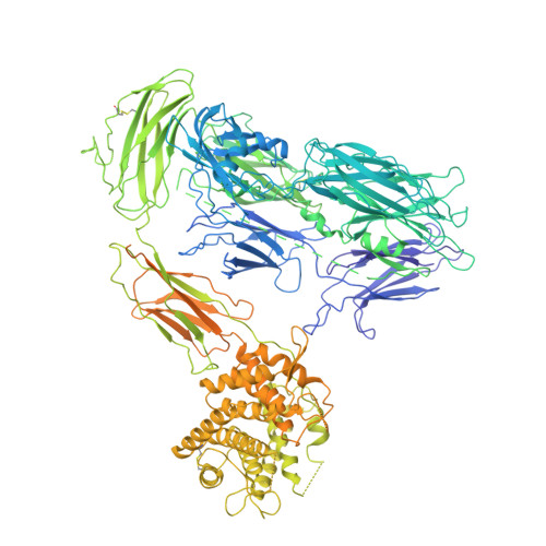
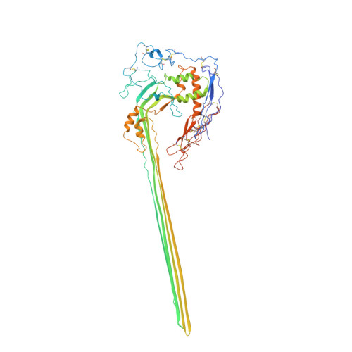
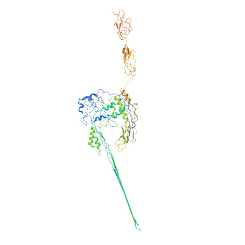
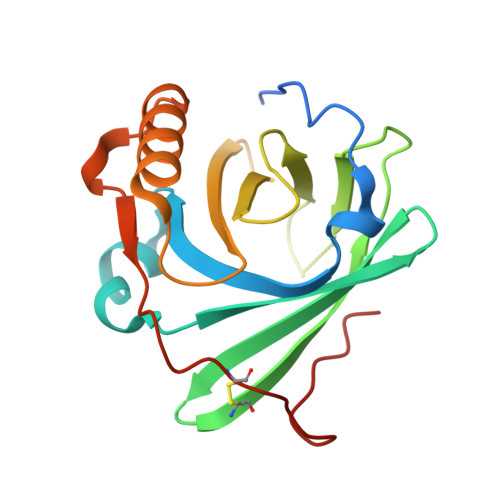
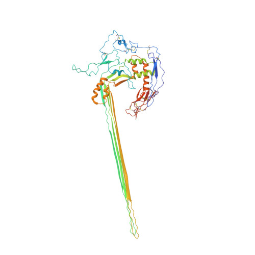
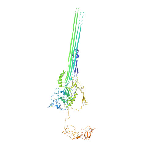
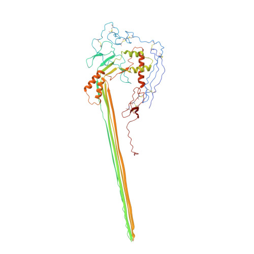
CryoEM reveals how the complement membrane attack complex ruptures lipid bilayers.

Menny, A.Serna, M.Boyd, C.M.Gardner, S.Joseph, A.P.Morgan, B.P.Topf, M.Brooks, N.J.Bubeck, D.

(2018) Nat Commun 9: 5316-5316

  • DOI: https://doi.org/10.1038/s41467-018-07653-5
  • Primary Citation of Related Structures:  
    6H03, 6H04

  • PubMed Abstract: 

    The membrane attack complex (MAC) is one of the immune system's first responders. Complement proteins assemble on target membranes to form pores that lyse pathogens and impact tissue homeostasis of self-cells. How MAC disrupts the membrane barrier remains unclear. Here we use electron cryo-microscopy and flicker spectroscopy to show that MAC interacts with lipid bilayers in two distinct ways. Whereas C6 and C7 associate with the outer leaflet and reduce the energy for membrane bending, C8 and C9 traverse the bilayer increasing membrane rigidity. CryoEM reconstructions reveal plasticity of the MAC pore and demonstrate how C5b6 acts as a platform, directing assembly of a giant β-barrel whose structure is supported by a glycan scaffold. Our work provides a structural basis for understanding how β-pore forming proteins breach the membrane and reveals a mechanism for how MAC kills pathogens and regulates cell functions.


  • Organizational Affiliation

    Department of Life Sciences, Sir Ernst Chain Building, Imperial College London, London, SW7 2AZ, UK.


Macromolecules
Find similar proteins by:  (by identity cutoff)  |  3D Structure
Entity ID: 1
MoleculeChains Sequence LengthOrganismDetailsImage
Complement C5,Complement C51,580Homo sapiensMutation(s): 0 
Membrane Entity: Yes 
UniProt & NIH Common Fund Data Resources
Find proteins for P01031 (Homo sapiens)
Explore P01031 
Go to UniProtKB:  P01031
PHAROS:  P01031
GTEx:  ENSG00000106804 
Entity Groups  
Sequence Clusters30% Identity50% Identity70% Identity90% Identity95% Identity100% Identity
UniProt GroupP01031
Sequence Annotations
Expand
  • Reference Sequence
Find similar proteins by:  (by identity cutoff)  |  3D Structure
Entity ID: 2
MoleculeChains Sequence LengthOrganismDetailsImage
Complement component C8 beta chainB [auth C]537Homo sapiensMutation(s): 0 
Membrane Entity: Yes 
UniProt & NIH Common Fund Data Resources
Find proteins for P07358 (Homo sapiens)
Explore P07358 
Go to UniProtKB:  P07358
PHAROS:  P07358
GTEx:  ENSG00000021852 
Entity Groups  
Sequence Clusters30% Identity50% Identity70% Identity90% Identity95% Identity100% Identity
UniProt GroupP07358
Sequence Annotations
Expand
  • Reference Sequence
Find similar proteins by:  (by identity cutoff)  |  3D Structure
Entity ID: 3
MoleculeChains Sequence LengthOrganismDetailsImage
Complement component C7C [auth D]821Homo sapiensMutation(s): 0 
Membrane Entity: Yes 
UniProt & NIH Common Fund Data Resources
Find proteins for P10643 (Homo sapiens)
Explore P10643 
Go to UniProtKB:  P10643
PHAROS:  P10643
GTEx:  ENSG00000112936 
Entity Groups  
Sequence Clusters30% Identity50% Identity70% Identity90% Identity95% Identity100% Identity
UniProt GroupP10643
Sequence Annotations
Expand
  • Reference Sequence
Find similar proteins by:  (by identity cutoff)  |  3D Structure
Entity ID: 4
MoleculeChains Sequence LengthOrganismDetailsImage
Complement component C8 gamma chainD [auth E]182Homo sapiensMutation(s): 0 
Membrane Entity: Yes 
UniProt & NIH Common Fund Data Resources
Find proteins for P07360 (Homo sapiens)
Explore P07360 
Go to UniProtKB:  P07360
PHAROS:  P07360
GTEx:  ENSG00000176919 
Entity Groups  
Sequence Clusters30% Identity50% Identity70% Identity90% Identity95% Identity100% Identity
UniProt GroupP07360
Sequence Annotations
Expand
  • Reference Sequence
Find similar proteins by:  (by identity cutoff)  |  3D Structure
Entity ID: 5
MoleculeChains Sequence LengthOrganismDetailsImage
Complement component C8 alpha chainE [auth F]554Homo sapiensMutation(s): 0 
Membrane Entity: Yes 
UniProt & NIH Common Fund Data Resources
Find proteins for P07357 (Homo sapiens)
Explore P07357 
Go to UniProtKB:  P07357
PHAROS:  P07357
GTEx:  ENSG00000157131 
Entity Groups  
Sequence Clusters30% Identity50% Identity70% Identity90% Identity95% Identity100% Identity
UniProt GroupP07357
Sequence Annotations
Expand
  • Reference Sequence
Find similar proteins by:  (by identity cutoff)  |  3D Structure
Entity ID: 6
MoleculeChains Sequence LengthOrganismDetailsImage
Complement component C6F [auth B]913Homo sapiensMutation(s): 0 
Membrane Entity: Yes 
UniProt & NIH Common Fund Data Resources
Find proteins for P13671 (Homo sapiens)
Explore P13671 
Go to UniProtKB:  P13671
PHAROS:  P13671
GTEx:  ENSG00000039537 
Entity Groups  
Sequence Clusters30% Identity50% Identity70% Identity90% Identity95% Identity100% Identity
UniProt GroupP13671
Sequence Annotations
Expand
  • Reference Sequence
Find similar proteins by:  (by identity cutoff)  |  3D Structure
Entity ID: 7
MoleculeChains Sequence LengthOrganismDetailsImage
Complement component C9538Homo sapiensMutation(s): 0 
Membrane Entity: Yes 
UniProt & NIH Common Fund Data Resources
Find proteins for P02748 (Homo sapiens)
Explore P02748 
Go to UniProtKB:  P02748
PHAROS:  P02748
GTEx:  ENSG00000113600 
Entity Groups  
Sequence Clusters30% Identity50% Identity70% Identity90% Identity95% Identity100% Identity
UniProt GroupP02748
Sequence Annotations
Expand
  • Reference Sequence
Oligosaccharides

Help

Entity ID: 8
MoleculeChains Length2D Diagram Glycosylation3D Interactions
beta-D-mannopyranose-(1-4)-2-acetamido-2-deoxy-beta-D-glucopyranose-(1-4)-2-acetamido-2-deoxy-beta-D-glucopyranose
Y
3N-Glycosylation
Glycosylation Resources
GlyTouCan:  G15407YE
GlyCosmos:  G15407YE
GlyGen:  G15407YE
Small Molecules
Ligands 1 Unique
IDChains Name / Formula / InChI Key2D Diagram3D Interactions
NAG
Query on NAG

Download Ideal Coordinates CCD File 
AB [auth I]
AC [auth W]
BB [auth I]
BC [auth W]
CB [auth J]
AB [auth I],
AC [auth W],
BB [auth I],
BC [auth W],
CB [auth J],
CC [auth X],
DB [auth J],
DC [auth X],
EB [auth K],
FB [auth K],
GB [auth L],
HB [auth L],
IB [auth M],
JB [auth M],
KB [auth N],
LB [auth N],
MB [auth O],
NB [auth O],
OB [auth Q],
PB [auth Q],
QB [auth R],
RB [auth R],
SB [auth S],
TA [auth F],
TB [auth S],
UA [auth G],
UB [auth T],
VA [auth G],
VB [auth T],
WA [auth P],
WB [auth U],
XA [auth P],
XB [auth U],
YA [auth H],
YB [auth V],
ZA [auth H],
ZB [auth V]
2-acetamido-2-deoxy-beta-D-glucopyranose
C8 H15 N O6
OVRNDRQMDRJTHS-FMDGEEDCSA-N
Experimental Data & Validation

Experimental Data

  • Method: ELECTRON MICROSCOPY
  • Resolution: 5.60 Å
  • Aggregation State: PARTICLE 
  • Reconstruction Method: SINGLE PARTICLE 
EM Software:
TaskSoftware PackageVersion
RECONSTRUCTIONRELION
MODEL REFINEMENTPHENIX

Structure Validation

View Full Validation Report


Entry History & Funding Information

Deposition Data


Funding OrganizationLocationGrant Number
Cancer Research UKUnited KingdomC26409/A16099
Medical Research Council (United Kingdom)United KingdomMR/M019292/1

Revision History  (Full details and data files)

  • Version 1.0: 2018-12-19
    Type: Initial release
  • Version 1.1: 2018-12-26
    Changes: Author supporting evidence, Data collection, Database references
  • Version 1.2: 2019-12-11
    Changes: Other
  • Version 2.0: 2020-07-29
    Type: Remediation
    Reason: Carbohydrate remediation
    Changes: Advisory, Atomic model, Data collection, Derived calculations, Structure summary