6GHM

Structure of PP1 alpha phosphatase bound to ASPP2


Experimental Data Snapshot

  • Method: X-RAY DIFFRACTION
  • Resolution: 2.15 Å
  • R-Value Free: 0.214 
  • R-Value Work: 0.177 
  • R-Value Observed: 0.179 

wwPDB Validation   3D Report Full Report


This is version 1.1 of the entry. See complete history


Literature

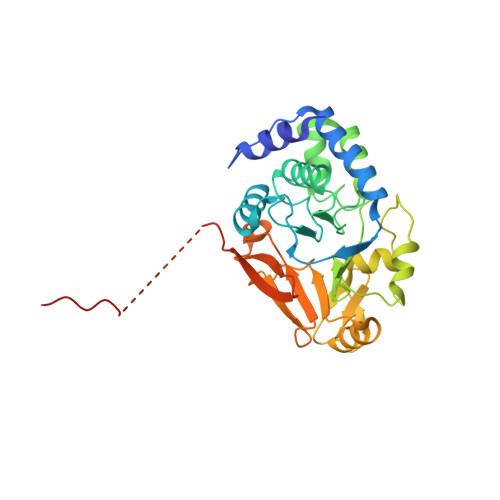
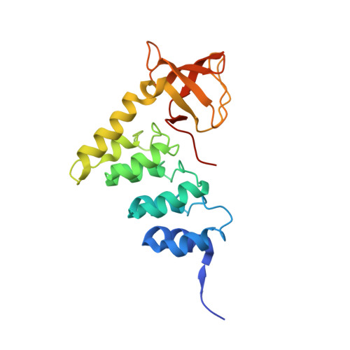
ASPP proteins discriminate between PP1 catalytic subunits through their SH3 domain and the PP1 C-tail.

Bertran, M.T.Mouilleron, S.Zhou, Y.Bajaj, R.Uliana, F.Kumar, G.S.van Drogen, A.Lee, R.Banerjee, J.J.Hauri, S.O'Reilly, N.Gstaiger, M.Page, R.Peti, W.Tapon, N.

(2019) Nat Commun 10: 771-771

  • DOI: https://doi.org/10.1038/s41467-019-08686-0
  • Primary Citation of Related Structures:  
    6GHM

  • PubMed Abstract: 

    Serine/threonine phosphatases such as PP1 lack substrate specificity and associate with a large array of targeting subunits to achieve the requisite selectivity. The tumour suppressor ASPP (apoptosis-stimulating protein of p53) proteins associate with PP1 catalytic subunits and are implicated in multiple functions from transcriptional regulation to cell junction remodelling. Here we show that Drosophila ASPP is part of a multiprotein PP1 complex and that PP1 association is necessary for several in vivo functions of Drosophila ASPP. We solve the crystal structure of the human ASPP2/PP1 complex and show that ASPP2 recruits PP1 using both its canonical RVxF motif, which binds the PP1 catalytic domain, and its SH3 domain, which engages the PP1 C-terminal tail. The ASPP2 SH3 domain can discriminate between PP1 isoforms using an acidic specificity pocket in the n-Src domain, providing an exquisite mechanism where multiple motifs are used combinatorially to tune binding affinity to PP1.


  • Organizational Affiliation

    Apoptosis and Proliferation Control Laboratory, The Francis Crick Institute, 1 Midland Road, London, NW1 1AT, UK.


Macromolecules
Find similar proteins by:  (by identity cutoff)  |  3D Structure
Entity ID: 1
MoleculeChains Sequence LengthOrganismDetailsImage
Serine/threonine-protein phosphatase PP1-alpha catalytic subunit
A, B
329Homo sapiensMutation(s): 0 
Gene Names: PPP1CAPPP1A
EC: 3.1.3.16
UniProt & NIH Common Fund Data Resources
Find proteins for P62136 (Homo sapiens)
Explore P62136 
Go to UniProtKB:  P62136
PHAROS:  P62136
GTEx:  ENSG00000172531 
Entity Groups  
Sequence Clusters30% Identity50% Identity70% Identity90% Identity95% Identity100% Identity
UniProt GroupP62136
Sequence Annotations
Expand
  • Reference Sequence
Find similar proteins by:  (by identity cutoff)  |  3D Structure
Entity ID: 2
MoleculeChains Sequence LengthOrganismDetailsImage
Apoptosis-stimulating of p53 protein 2
C, D
214Homo sapiensMutation(s): 0 
Gene Names: TP53BP2ASPP2BBP
UniProt & NIH Common Fund Data Resources
Find proteins for Q13625 (Homo sapiens)
Explore Q13625 
Go to UniProtKB:  Q13625
PHAROS:  Q13625
GTEx:  ENSG00000143514 
Entity Groups  
Sequence Clusters30% Identity50% Identity70% Identity90% Identity95% Identity100% Identity
UniProt GroupQ13625
Sequence Annotations
Expand
  • Reference Sequence
Small Molecules
Ligands 4 Unique
IDChains Name / Formula / InChI Key2D Diagram3D Interactions
NHE
Query on NHE

Download Ideal Coordinates CCD File 
DA [auth D]
N [auth A]
O [auth A]
W [auth B]
X [auth B]
DA [auth D],
N [auth A],
O [auth A],
W [auth B],
X [auth B],
Y [auth B]
2-[N-CYCLOHEXYLAMINO]ETHANE SULFONIC ACID
C8 H17 N O3 S
MKWKNSIESPFAQN-UHFFFAOYSA-N
GOL
Query on GOL

Download Ideal Coordinates CCD File 
K [auth A],
L [auth A],
M [auth A],
V [auth B]
GLYCEROL
C3 H8 O3
PEDCQBHIVMGVHV-UHFFFAOYSA-N
EDO
Query on EDO

Download Ideal Coordinates CCD File 
BA [auth D]
CA [auth D]
E [auth A]
F [auth A]
G [auth A]
BA [auth D],
CA [auth D],
E [auth A],
F [auth A],
G [auth A],
H [auth A],
I [auth A],
J [auth A],
R [auth B],
S [auth B],
T [auth B],
U [auth B]
1,2-ETHANEDIOL
C2 H6 O2
LYCAIKOWRPUZTN-UHFFFAOYSA-N
MN
Query on MN

Download Ideal Coordinates CCD File 
AA [auth B],
P [auth A],
Q [auth A],
Z [auth B]
MANGANESE (II) ION
Mn
WAEMQWOKJMHJLA-UHFFFAOYSA-N
Experimental Data & Validation

Experimental Data

  • Method: X-RAY DIFFRACTION
  • Resolution: 2.15 Å
  • R-Value Free: 0.214 
  • R-Value Work: 0.177 
  • R-Value Observed: 0.179 
  • Space Group: P 1
Unit Cell:
Length ( Å )Angle ( ˚ )
a = 46.84α = 91.06
b = 81.624β = 91.84
c = 87.966γ = 103.89
Software Package:
Software NamePurpose
PHENIXrefinement
DIALSdata reduction
Aimlessdata scaling
PHASERphasing

Structure Validation

View Full Validation Report


Entry History 

Deposition Data

Revision History  (Full details and data files)

  • Version 1.0: 2019-02-27
    Type: Initial release
  • Version 1.1: 2024-01-17
    Changes: Data collection, Database references, Derived calculations, Refinement description