6GEL

The structure of TWITCH-2B


Experimental Data Snapshot

  • Method: X-RAY DIFFRACTION
  • Resolution: 2.51 Å
  • R-Value Free: 0.240 
  • R-Value Work: 0.195 
  • R-Value Observed: 0.198 

wwPDB Validation   3D Report Full Report


This is version 1.2 of the entry. See complete history


Literature

Dynamic tuning of FRET in a green fluorescent protein biosensor.

Trigo-Mourino, P.Thestrup, T.Griesbeck, O.Griesinger, C.Becker, S.

(2019) Sci Adv 5: eaaw4988-eaaw4988

  • DOI: https://doi.org/10.1126/sciadv.aaw4988
  • Primary Citation of Related Structures:  
    6GEL, 6GEZ

  • PubMed Abstract: 

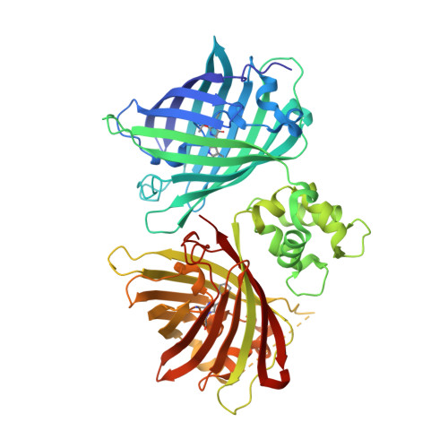
    Förster resonance energy transfer (FRET) between mutants of green fluorescent protein is widely used to monitor protein-protein interactions and as a readout mode in fluorescent biosensors. Despite the fundamental importance of distance and molecular angles of fluorophores to each other, structural details on fluorescent protein FRET have been missing. Here, we report the high-resolution x-ray structure of the fluorescent proteins mCerulean3 and cpVenus within the biosensor Twitch-2B, as they undergo FRET and characterize the dynamics of this biosensor with B 0 2 -dependent paramagnetic nuclear magnetic resonance at 900 MHz and 1.1 GHz. These structural data provide the unprecedented opportunity to calculate FRET from the x-ray structure and to compare it to experimental data in solution. We find that interdomain dynamics limits the FRET effect and show that a rigidification of the sensor further enhances FRET.


  • Organizational Affiliation

    Department for NMR-Based Structural Biology, Max Planck Institute for Biophysical Chemistry, Göttingen, Germany.


Macromolecules
Find similar proteins by:  (by identity cutoff)  |  3D Structure
Entity ID: 1
MoleculeChains Sequence LengthOrganismDetailsImage
Green fluorescent protein,Optimized Ratiometric Calcium Sensor,Green fluorescent protein,Green fluorescent protein
A, B
552Aequorea victoriaOpsanus tauMutation(s): 2 
Gene Names: GFP
UniProt
Find proteins for P42212 (Aequorea victoria)
Explore P42212 
Go to UniProtKB:  P42212
Find proteins for W5IDB2 (Opsanus tau)
Explore W5IDB2 
Go to UniProtKB:  W5IDB2
Entity Groups  
Sequence Clusters30% Identity50% Identity70% Identity90% Identity95% Identity100% Identity
UniProt GroupsP42212W5IDB2
Sequence Annotations
Expand
  • Reference Sequence
Small Molecules
Ligands 4 Unique
IDChains Name / Formula / InChI Key2D Diagram3D Interactions
PG4
Query on PG4

Download Ideal Coordinates CCD File 
F [auth A],
G [auth A]
TETRAETHYLENE GLYCOL
C8 H18 O5
UWHCKJMYHZGTIT-UHFFFAOYSA-N
GOL
Query on GOL

Download Ideal Coordinates CCD File 
E [auth A],
L [auth B],
M [auth B]
GLYCEROL
C3 H8 O3
PEDCQBHIVMGVHV-UHFFFAOYSA-N
FMT
Query on FMT

Download Ideal Coordinates CCD File 
H [auth A],
I [auth A],
N [auth B],
O [auth B],
P [auth B]
FORMIC ACID
C H2 O2
BDAGIHXWWSANSR-UHFFFAOYSA-N
CA
Query on CA

Download Ideal Coordinates CCD File 
C [auth A],
D [auth A],
J [auth B],
K [auth B]
CALCIUM ION
Ca
BHPQYMZQTOCNFJ-UHFFFAOYSA-N
Modified Residues  2 Unique
IDChains TypeFormula2D DiagramParent
CR2
Query on CR2
A, B
L-PEPTIDE LINKINGC13 H13 N3 O4GLY, TYR, GLY
CRF
Query on CRF
A, B
L-PEPTIDE LINKINGC17 H18 N4 O4THR, TRP, GLY
Experimental Data & Validation

Experimental Data

  • Method: X-RAY DIFFRACTION
  • Resolution: 2.51 Å
  • R-Value Free: 0.240 
  • R-Value Work: 0.195 
  • R-Value Observed: 0.198 
  • Space Group: P 21 21 21
Unit Cell:
Length ( Å )Angle ( ˚ )
a = 58.075α = 90
b = 156.773β = 90
c = 169.284γ = 90
Software Package:
Software NamePurpose
REFMACrefinement
PDB_EXTRACTdata extraction
XDSdata reduction
SADABSdata scaling
AutoSolphasing

Structure Validation

View Full Validation Report


Entry History 

Deposition Data

Revision History  (Full details and data files)

  • Version 1.0: 2019-08-21
    Type: Initial release
  • Version 1.1: 2019-09-04
    Changes: Data collection, Database references
  • Version 1.2: 2019-09-11
    Changes: Data collection, Database references