6D26

Crystal structure of the prostaglandin D2 receptor CRTH2 with fevipiprant


Experimental Data Snapshot

  • Method: X-RAY DIFFRACTION
  • Resolution: 2.80 Å
  • R-Value Free: 0.280 
  • R-Value Work: 0.250 
  • R-Value Observed: 0.252 

wwPDB Validation   3D Report Full Report


Ligand Structure Quality Assessment 


This is version 1.3 of the entry. See complete history


Literature

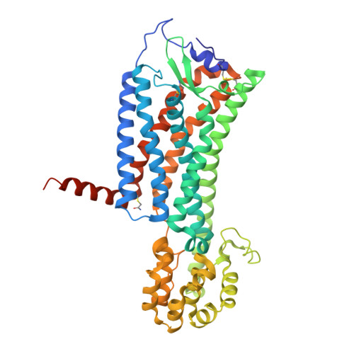
Structures of the Human PGD2Receptor CRTH2 Reveal Novel Mechanisms for Ligand Recognition.

Wang, L.Yao, D.Deepak, R.N.V.K.Liu, H.Xiao, Q.Fan, H.Gong, W.Wei, Z.Zhang, C.

(2018) Mol Cell 72: 48-59.e4

  • DOI: https://doi.org/10.1016/j.molcel.2018.08.009
  • Primary Citation of Related Structures:  
    6D26, 6D27

  • PubMed Abstract: 

    The signaling of prostaglandin D 2 (PGD 2 ) through G-protein-coupled receptor (GPCR) CRTH2 is a major pathway in type 2 inflammation. Compelling evidence suggests the therapeutic benefits of blocking CRTH2 signaling in many inflammatory disorders. Currently, a number of CRTH2 antagonists are under clinical investigation, and one compound, fevipiprant, has advanced to phase 3 clinical trials for asthma. Here, we present the crystal structures of human CRTH2 with two antagonists, fevipiprant and CAY10471. The structures, together with docking and ligand-binding data, reveal a semi-occluded pocket covered by a well-structured amino terminus and different binding modes of chemically diverse CRTH2 antagonists. Structural analysis suggests a ligand entry port and a binding process that is facilitated by opposite charge attraction for PGD 2 , which differs significantly from the binding pose and binding environment of lysophospholipids and endocannabinoids, revealing a new mechanism for lipid recognition by GPCRs.


  • Organizational Affiliation

    Department of Pharmacology and Chemical Biology, School of Medicine, University of Pittsburgh, Pittsburgh, PA 15261, USA.


Macromolecules
Find similar proteins by:  (by identity cutoff)  |  3D Structure
Entity ID: 1
MoleculeChains Sequence LengthOrganismDetailsImage
Prostaglandin D2 receptor 2, Endolysin chimera470Homo sapiensTequatrovirus T4Mutation(s): 3 
Gene Names: PTGDR2CRTH2DL1RGPR44eT4Tp126
Membrane Entity: Yes 
UniProt & NIH Common Fund Data Resources
Find proteins for Q9Y5Y4 (Homo sapiens)
Explore Q9Y5Y4 
Go to UniProtKB:  Q9Y5Y4
PHAROS:  Q9Y5Y4
GTEx:  ENSG00000183134 
Find proteins for P00720 (Enterobacteria phage T4)
Explore P00720 
Go to UniProtKB:  P00720
Entity Groups  
Sequence Clusters30% Identity50% Identity70% Identity90% Identity95% Identity100% Identity
UniProt GroupsP00720Q9Y5Y4
Sequence Annotations
Expand
  • Reference Sequence
Small Molecules
Ligands 6 Unique
IDChains Name / Formula / InChI Key2D Diagram3D Interactions
FSY
Query on FSY

Download Ideal Coordinates CCD File 
B [auth A]fevipiprant
C19 H17 F3 N2 O4 S
GFPPXZDRVCSVNR-UHFFFAOYSA-N
OLA
Query on OLA

Download Ideal Coordinates CCD File 
R [auth A]OLEIC ACID
C18 H34 O2
ZQPPMHVWECSIRJ-KTKRTIGZSA-N
PGE
Query on PGE

Download Ideal Coordinates CCD File 
Q [auth A]TRIETHYLENE GLYCOL
C6 H14 O4
ZIBGPFATKBEMQZ-UHFFFAOYSA-N
SIN
Query on SIN

Download Ideal Coordinates CCD File 
L [auth A],
M [auth A],
N [auth A],
O [auth A],
P [auth A]
SUCCINIC ACID
C4 H6 O4
KDYFGRWQOYBRFD-UHFFFAOYSA-N
SO4
Query on SO4

Download Ideal Coordinates CCD File 
C [auth A]
D [auth A]
E [auth A]
F [auth A]
G [auth A]
C [auth A],
D [auth A],
E [auth A],
F [auth A],
G [auth A],
H [auth A],
I [auth A],
J [auth A],
K [auth A]
SULFATE ION
O4 S
QAOWNCQODCNURD-UHFFFAOYSA-L
PGO
Query on PGO

Download Ideal Coordinates CCD File 
S [auth A]S-1,2-PROPANEDIOL
C3 H8 O2
DNIAPMSPPWPWGF-VKHMYHEASA-N
Modified Residues  1 Unique
IDChains TypeFormula2D DiagramParent
YCM
Query on YCM
A
L-PEPTIDE LINKINGC5 H10 N2 O3 SCYS
Binding Affinity Annotations 
IDSourceBinding Affinity
FSY BindingDB:  6D26 Ki: 4 (nM) from 1 assay(s)
Kd: 1 (nM) from 1 assay(s)
IC50: min: 0.4, max: 3.1 (nM) from 4 assay(s)
Experimental Data & Validation

Experimental Data

  • Method: X-RAY DIFFRACTION
  • Resolution: 2.80 Å
  • R-Value Free: 0.280 
  • R-Value Work: 0.250 
  • R-Value Observed: 0.252 
  • Space Group: P 21 21 21
Unit Cell:
Length ( Å )Angle ( ˚ )
a = 50.457α = 90
b = 61.71β = 90
c = 266.517γ = 90
Software Package:
Software NamePurpose
PHENIXrefinement
HKL-2000data scaling
PHASERphasing
PDB_EXTRACTdata extraction
HKL-2000data reduction

Structure Validation

View Full Validation Report


Ligand Structure Quality Assessment 


Entry History & Funding Information

Deposition Data


Funding OrganizationLocationGrant Number
National Natural Science Foundation of China (NSFC)China--

Revision History  (Full details and data files)

  • Version 1.0: 2018-10-03
    Type: Initial release
  • Version 1.1: 2019-04-17
    Changes: Data collection, Database references
  • Version 1.2: 2020-01-08
    Changes: Author supporting evidence
  • Version 1.3: 2023-10-04
    Changes: Data collection, Database references, Refinement description