6CYQ

Crystal structure of CTX-M-14 S70G/N106S beta-lactamase in complex with hydrolyzed cefotaxime


Experimental Data Snapshot

  • Method: X-RAY DIFFRACTION
  • Resolution: 1.70 Å
  • R-Value Free: 0.200 
  • R-Value Work: 0.170 
  • R-Value Observed: 0.172 

wwPDB Validation   3D Report Full Report


Ligand Structure Quality Assessment 


This is version 1.5 of the entry. See complete history


Literature

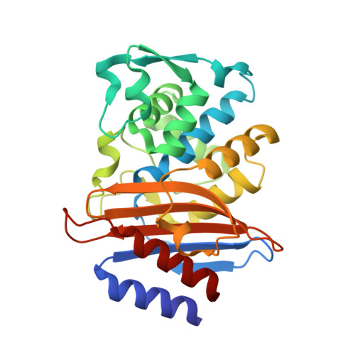
Synergistic effects of functionally distinct substitutions in beta-lactamase variants shed light on the evolution of bacterial drug resistance.

Patel, M.P.Hu, L.Brown, C.A.Sun, Z.Adamski, C.J.Stojanoski, V.Sankaran, B.Prasad, B.V.V.Palzkill, T.

(2018) J Biol Chem 293: 17971-17984

  • DOI: https://doi.org/10.1074/jbc.RA118.003792
  • Primary Citation of Related Structures:  
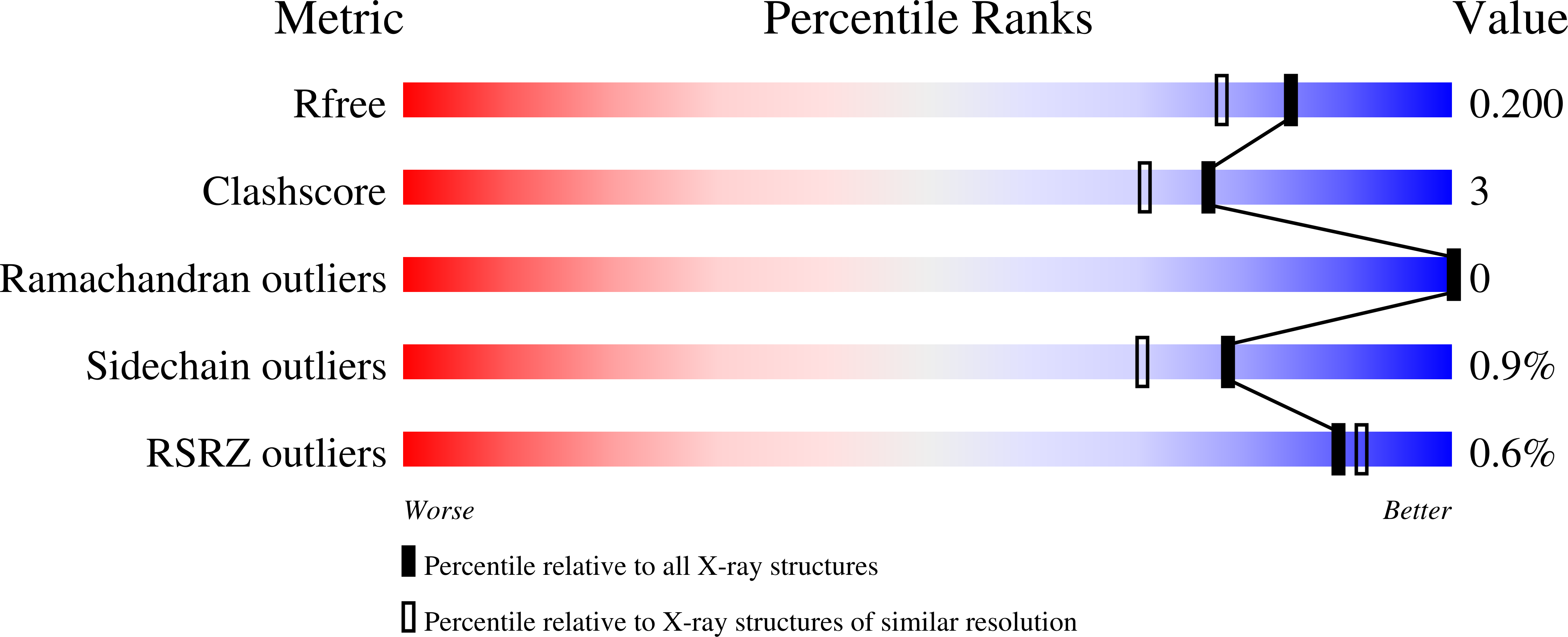
    6CYK, 6CYN, 6CYQ, 6CYU

  • PubMed Abstract: 

    The CTX-M β-lactamases have emerged as the most widespread extended-spectrum β-lactamases (ESBLs) in Gram-negative bacteria. These enzymes rapidly hydrolyze cefotaxime, but not the related cephalosporin, ceftazidime. ESBL variants have evolved, however, that provide enhanced ceftazidime resistance. We show here that a natural variant at a nonactive site, i.e. second-shell residue N106S, enhances enzyme stability but reduces catalytic efficiency for cefotaxime and ceftazidime and decreases resistance levels. However, when the N106S variant was combined with an active-site variant, D240G, that enhances enzyme catalytic efficiency, but decreases stability, the resultant double mutant exhibited higher resistance levels than predicted on the basis of the phenotypes of each variant. We found that this epistasis is due to compensatory effects, whereby increased stability provided by N106S overrides its cost of decreased catalytic activity. X-ray structures of the variant enzymes in complex with cefotaxime revealed conformational changes in the active-site loop spanning residues 103-106 that were caused by the N106S substitution and relieve steric strain to stabilize the enzyme, but also alter contacts with cefotaxime and thereby reduce catalytic activity. We noted that the 103-106 loop conformation in the N106S-containing variants is different from that of WT CTX-M but nearly identical to that of the non-ESBL, TEM-1 β-lactamase, having a serine at the 106 position. Therefore, residue 106 may serve as a "switch" that toggles the conformations of the 103-106 loop. When it is serine, the loop is in the non-ESBL, TEM-like conformation, and when it is asparagine, the loop is in a CTX-M-like, cefotaximase-favorable conformation.


  • Organizational Affiliation

    From the Interdepartmental Graduate Program in Translational Biology and Molecular Medicine; Department of Pharmacology and Chemical Biology, Baylor College of Medicine, Houston, Texas 77030.


Macromolecules
Find similar proteins by:  (by identity cutoff)  |  3D Structure
Entity ID: 1
MoleculeChains Sequence LengthOrganismDetailsImage
Beta-lactamase
A, B, C, D, E
A, B, C, D, E, F, G, H
263Escherichia coliMutation(s): 2 
Gene Names: 
EC: 3.5.2.6
UniProt
Find proteins for Q9L5C8 (Escherichia coli)
Explore Q9L5C8 
Go to UniProtKB:  Q9L5C8
Entity Groups  
Sequence Clusters30% Identity50% Identity70% Identity90% Identity95% Identity100% Identity
UniProt GroupQ9L5C8
Sequence Annotations
Expand
  • Reference Sequence
Small Molecules
Ligands 2 Unique
IDChains Name / Formula / InChI Key2D Diagram3D Interactions
CE4
Query on CE4

Download Ideal Coordinates CCD File 
I [auth A]
J [auth B]
K [auth C]
L [auth D]
M [auth E]
I [auth A],
J [auth B],
K [auth C],
L [auth D],
M [auth E],
N [auth F],
P [auth G],
Q [auth H]
(2R)-2-[(R)-{[(2Z)-2-(2-amino-1,3-thiazol-4-yl)-2-(methoxyimino)acetyl]amino}(carboxy)methyl]-5-methylidene-5,6-dihydro -2H-1,3-thiazine-4-carboxylic acid
C14 H15 N5 O6 S2
ZRDXEERHKBZJMY-KTFFUYHOSA-N
GOL
Query on GOL

Download Ideal Coordinates CCD File 
O [auth F]GLYCEROL
C3 H8 O3
PEDCQBHIVMGVHV-UHFFFAOYSA-N
Experimental Data & Validation

Experimental Data

  • Method: X-RAY DIFFRACTION
  • Resolution: 1.70 Å
  • R-Value Free: 0.200 
  • R-Value Work: 0.170 
  • R-Value Observed: 0.172 
  • Space Group: P 32
Unit Cell:
Length ( Å )Angle ( ˚ )
a = 83.375α = 90
b = 83.375β = 90
c = 232.058γ = 120
Software Package:
Software NamePurpose
PHENIXrefinement
HKL-2000data reduction
HKL-2000data scaling
PHASERphasing

Structure Validation

View Full Validation Report


Ligand Structure Quality Assessment 


Entry History & Funding Information

Deposition Data


Funding OrganizationLocationGrant Number
National Institutes of Health/National Institute Of Allergy and Infectious Diseases (NIH/NIAID)United StatesR01 AI32956

Revision History  (Full details and data files)

  • Version 1.0: 2018-10-10
    Type: Initial release
  • Version 1.1: 2018-10-17
    Changes: Data collection, Database references, Structure summary
  • Version 1.2: 2018-11-28
    Changes: Data collection, Database references
  • Version 1.3: 2019-12-18
    Changes: Author supporting evidence
  • Version 1.4: 2021-06-30
    Changes: Derived calculations, Structure summary
  • Version 1.5: 2023-10-04
    Changes: Data collection, Database references, Refinement description