Ab initio structure solution of a proteolytic fragment using ARCIMBOLDO.
Abendroth, J., Sankaran, B., Myler, P.J., Lorimer, D.D., Edwards, T.E.(2018) Acta Crystallogr F Struct Biol Commun 74: 530-535
- PubMed: 30198884
- DOI: https://doi.org/10.1107/S2053230X18010063
- Primary Citation of Related Structures:
6CUM - PubMed Abstract:
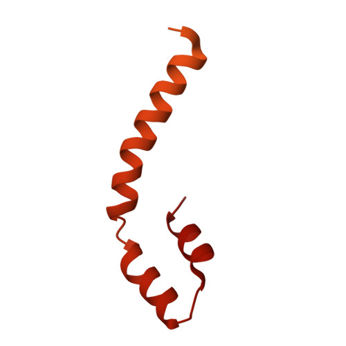
Crystal structure determination requires solving the phase problem. This can be accomplished using ab initio direct methods for small molecules and macromolecules at resolutions higher than 1.2 Å, whereas macromolecular structure determination at lower resolution requires either molecular replacement using a homologous structure or experimental phases using a derivative such as covalent labeling (for example selenomethionine or mercury derivatization) or heavy-atom soaking (for example iodide ions). Here, a case is presented in which crystals were obtained from a 30.8 kDa protein sample and yielded a 1.6 Å resolution data set with a unit cell that could accommodate approximately 8 kDa of protein. Thus, it was unclear what had been crystallized. Molecular replacement with pieces of homologous proteins and attempts at iodide ion soaking failed to yield a solution. The crystals could not be reproduced. Sequence-independent molecular replacement using the structures available in the Protein Data Bank also failed to yield a solution. Ultimately, ab initio structure solution proved successful using the program ARCIMBOLDO, which identified two α-helical elements and yielded interpretable maps. The structure was the C-terminal dimerization domain of the intended target from Mycobacterium smegmatis. This structure is presented as a user-friendly test case in which an unknown protein fragment could be determined using ARCIMBOLDO.
Organizational Affiliation:
Seattle Structural Genomics Center for Infectious Disease (SSGCID), Seattle, Washington, USA.