Crystal structures of a novel family IV esterase in free and substrate-bound form.
Hoppner, A., Bollinger, A., Kobus, S., Thies, S., Coscolin, C., Ferrer, M., Jaeger, K.E., Smits, S.H.J.(2021) FEBS J 288: 3570-3584
- PubMed: 33342083
- DOI: https://doi.org/10.1111/febs.15680
- Primary Citation of Related Structures:
6Z68, 6Z69 - PubMed Abstract:
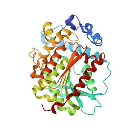
Bacterial lipolytic enzymes of family IV are homologs of the mammalian hormone-sensitive lipases (HSL) and have been successfully used for various biotechnological applications. The broad substrate specificity and ability for enantio-, regio-, and stereoselective hydrolysis are remarkable features of enzymes from this class. Many crystal structures are available for esterases and lipases, but structures of enzyme-substrate or enzyme-inhibitor complexes are less frequent although important to understand the molecular basis of enzyme-substrate interaction and to rationalize biochemical enzyme characteristics. Here, we report on the structures of a novel family IV esterase isolated from a metagenomic screen, which shows a broad substrate specificity. We solved the crystal structures in the apo form and with a bound substrate analogue at 1.35 and 1.81 Å resolution, respectively. This enzyme named PtEst1 hydrolyzed more than 60 out 96 structurally different ester substrates thus being substrate promiscuous. Its broad substrate specificity is in accord with a large active site cavity, which is covered by an α-helical cap domain. The substrate analogue methyl 4-methylumbelliferyl hexylphosphonate was rapidly hydrolyzed by the enzyme leading to a complete inactivation caused by covalent binding of phosphinic acid to the catalytic serine. Interestingly, the alcohol leaving group 4-methylumbelliferone was found remaining in the active site cavity, and additionally, a complete inhibitor molecule was found at the cap domain next to the entrance of the substrate tunnel. This unique situation allowed gaining valuable insights into the role of the cap domain for enzyme-substrate interaction of esterases belonging to family IV. DATABASE: Structural data of PtEst1 are available in the worldwide protein data bank (https://www.rcsb.org) under the accession codes: 6Z68 (apo-PtEst1) and 6Z69 (PtEst1-inhibitor complex).
Organizational Affiliation:
Center for Structural Studies, Heinrich-Heine-University, Düsseldorf, Germany.