6YV9

Mannosyltransferase PcManGT from Pyrobaculum calidifontis in complex with GDP-Man and Mn2+


Experimental Data Snapshot

  • Method: X-RAY DIFFRACTION
  • Resolution: 2.70 Å
  • R-Value Free: 0.265 
  • R-Value Work: 0.215 
  • R-Value Observed: 0.220 

wwPDB Validation   3D Report Full Report


Ligand Structure Quality Assessment 


This is version 1.1 of the entry. See complete history


Literature

A Transmembrane Crenarchaeal Mannosyltransferase Is Involved in N-Glycan Biosynthesis and Displays an Unexpected Minimal Cellulose-Synthase-like Fold.

Gandini, R.Reichenbach, T.Spadiut, O.Tan, T.C.Kalyani, D.C.Divne, C.

(2020) J Mol Biol 432: 4658-4672

  • DOI: https://doi.org/10.1016/j.jmb.2020.06.016
  • Primary Citation of Related Structures:  
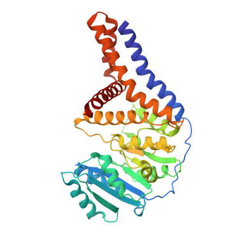
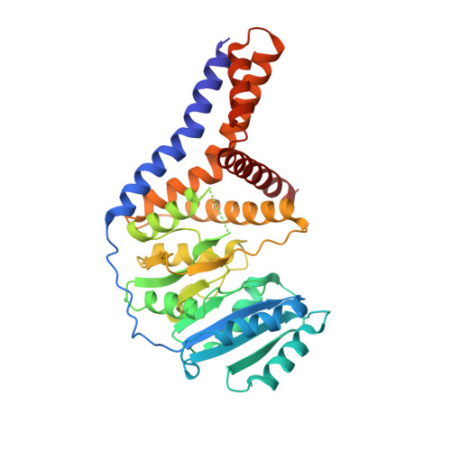
    6YV7, 6YV8, 6YV9

  • PubMed Abstract: 

    Protein glycosylation constitutes a critical post-translational modification that supports a vast number of biological functions in living organisms across all domains of life. A seemingly boundless number of enzymes, glycosyltransferases, are involved in the biosynthesis of these protein-linked glycans. Few glycan-biosynthetic glycosyltransferases have been characterized in vitro, mainly due to the majority being integral membrane proteins and the paucity of relevant acceptor substrates. The crenarchaeote Pyrobaculum calidifontis belongs to the TACK superphylum of archaea (Thaumarchaeota, Aigarchaeota, Crenarchaeota, Korarchaeota) that has been proposed as an eukaryotic ancestor. In archaea, N-glycans are mainly found on cell envelope surface-layer proteins, archaeal flagellins and pili. Archaeal N-glycans are distinct from those of eukaryotes, but one noteworthy exception is the high-mannose N-glycan produced by P. calidifontis, which is similar in sugar composition to the eukaryotic counterpart. Here, we present the characterization and crystal structure of the first member of a crenarchaeal membrane glycosyltransferase, PcManGT. We show that the enzyme is a GDP-, dolichylphosphate-, and manganese-dependent mannosyltransferase. The membrane domain of PcManGT includes three transmembrane helices that topologically coincide with "half" of the six-transmembrane helix cellulose-binding tunnel in Rhodobacter spheroides cellulose synthase BcsA. Conceivably, this "half tunnel" would be suitable for binding the dolichylphosphate-linked acceptor substrate. The PcManGT gene (Pcal_0472) is located in a large gene cluster comprising 14 genes of which 6 genes code for glycosyltransferases, and we hypothesize that this cluster may constitute a crenarchaeal N-glycosylation (PNG) gene cluster.


  • Organizational Affiliation

    School of Engineering Sciences in Chemistry, Biotechnology, and Health (CBH), KTH Royal Institute of Technology, 10691 Stockholm, Sweden.


Macromolecules
Find similar proteins by:  (by identity cutoff)  |  3D Structure
Entity ID: 1
MoleculeChains Sequence LengthOrganismDetailsImage
Glycosyl transferase, family 2355Pyrobaculum calidifontis JCM 11548Mutation(s): 0 
Gene Names: Pcal_0472
UniProt
Find proteins for A3MTD6 (Pyrobaculum calidifontis (strain DSM 21063 / JCM 11548 / VA1))
Explore A3MTD6 
Go to UniProtKB:  A3MTD6
Entity Groups  
Sequence Clusters30% Identity50% Identity70% Identity90% Identity95% Identity100% Identity
UniProt GroupA3MTD6
Sequence Annotations
Expand
  • Reference Sequence
Find similar proteins by:  (by identity cutoff)  |  3D Structure
Entity ID: 2
MoleculeChains Sequence LengthOrganismDetailsImage
Glycosyl transferase, family 2356Pyrobaculum calidifontis JCM 11548Mutation(s): 0 
Gene Names: Pcal_0472
UniProt
Find proteins for A3MTD6 (Pyrobaculum calidifontis (strain DSM 21063 / JCM 11548 / VA1))
Explore A3MTD6 
Go to UniProtKB:  A3MTD6
Entity Groups  
Sequence Clusters30% Identity50% Identity70% Identity90% Identity95% Identity100% Identity
UniProt GroupA3MTD6
Sequence Annotations
Expand
  • Reference Sequence
Experimental Data & Validation

Experimental Data

  • Method: X-RAY DIFFRACTION
  • Resolution: 2.70 Å
  • R-Value Free: 0.265 
  • R-Value Work: 0.215 
  • R-Value Observed: 0.220 
  • Space Group: P 21 21 21
Unit Cell:
Length ( Å )Angle ( ˚ )
a = 51.465α = 90
b = 107.418β = 90
c = 163.845γ = 90
Software Package:
Software NamePurpose
PHENIXrefinement
XDSdata reduction
XSCALEdata scaling
PHENIXphasing

Structure Validation

View Full Validation Report


Ligand Structure Quality Assessment 


Entry History & Funding Information

Deposition Data


Funding OrganizationLocationGrant Number
Swedish Research CouncilSweden2011-5768
Swedish Research CouncilSweden2013-5717
Swedish Research CouncilSweden2017-03877

Revision History  (Full details and data files)

  • Version 1.0: 2020-07-22
    Type: Initial release
  • Version 1.1: 2020-08-05
    Changes: Database references, Derived calculations