


Substrate Engagement and Catalytic Mechanisms of N-Acetylglucosaminyltransferase V
Darby, J.F., Gilio, A.K., Piniello, B., Roth, C., Blagova, E., Rovira, C., Hubbard, R.E., Davies, G.J., Wu, L.(2020) ACS Catal
Experimental Data Snapshot
(2020) ACS Catal
Entity ID: 1 | |||||
|---|---|---|---|---|---|
| Molecule | Chains | Sequence Length | Organism | Details | Image |
| Alpha-1,6-mannosylglycoprotein 6-beta-N-acetylglucosaminyltransferase A | A [auth AAA], B [auth BBB] | 515 | Homo sapiens | Mutation(s): 0 Gene Names: MGAT5, GGNT5 EC: 2.4.1.155 | 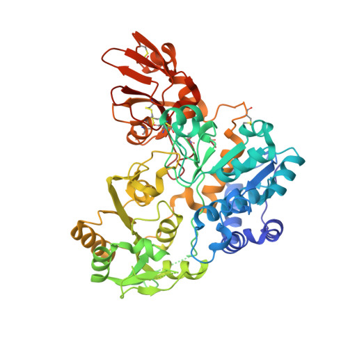 |
UniProt & NIH Common Fund Data Resources | |||||
Find proteins for Q09328 (Homo sapiens) Explore Q09328 Go to UniProtKB: Q09328 | |||||
PHAROS: Q09328 GTEx: ENSG00000152127 | |||||
Entity Groups | |||||
| Sequence Clusters | 30% Identity50% Identity70% Identity90% Identity95% Identity100% Identity | ||||
| UniProt Group | Q09328 | ||||
Sequence AnnotationsExpand | |||||
| |||||
| Ligands 3 Unique | |||||
|---|---|---|---|---|---|
| ID | Chains | Name / Formula / InChI Key | 2D Diagram | 3D Interactions | |
| U2F (Subject of Investigation/LOI) Query on U2F | F [auth AAA], K [auth BBB] | URIDINE-5'-DIPHOSPHATE-2-DEOXY-2-FLUORO-ALPHA-D-GLUCOSE C15 H23 F N2 O16 P2 NGTCPFGWXMBZEP-NQQHDEILSA-N |  | ||
| SO4 Query on SO4 | G [auth AAA] H [auth AAA] I [auth AAA] L [auth BBB] M [auth BBB] | SULFATE ION O4 S QAOWNCQODCNURD-UHFFFAOYSA-L |  | ||
| EDO Query on EDO | E [auth AAA], J [auth BBB] | 1,2-ETHANEDIOL C2 H6 O2 LYCAIKOWRPUZTN-UHFFFAOYSA-N |  | ||
| Length ( Å ) | Angle ( ˚ ) |
|---|---|
| a = 46.52 | α = 108.42 |
| b = 69.21 | β = 92.25 |
| c = 90.65 | γ = 106.54 |
| Software Name | Purpose |
|---|---|
| REFMAC | refinement |
| xia2 | data reduction |
| xia2 | data scaling |
| REFMAC | phasing |
| Funding Organization | Location | Grant Number |
|---|---|---|
| European Research Council (ERC) | United Kingdom | ErC-2012-AdG-322942 |
RCSB PDB (citation) is hosted by
RCSB PDB is a member of the


