6XRF

EagT6 Tse6 NT complex


Experimental Data Snapshot

  • Method: X-RAY DIFFRACTION
  • Resolution: 2.56 Å
  • R-Value Free: 0.268 
  • R-Value Work: 0.230 
  • R-Value Observed: 0.232 

wwPDB Validation   3D Report Full Report


This is version 1.1 of the entry. See complete history


Literature

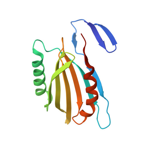
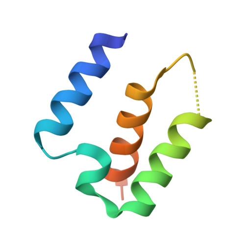
Structural basis for effector transmembrane domain recognition by type VI secretion system chaperones.

Ahmad, S.Tsang, K.K.Sachar, K.Quentin, D.Tashin, T.M.Bullen, N.P.Raunser, S.McArthur, A.G.Prehna, G.Whitney, J.C.

(2020) Elife 9

  • DOI: https://doi.org/10.7554/eLife.62816
  • Primary Citation of Related Structures:  
    6XRB, 6XRF, 6XRR

  • PubMed Abstract: 

    Type VI secretion systems (T6SSs) deliver antibacterial effector proteins between neighboring bacteria. Many effectors harbor N-terminal t rans m embrane d omains (TMDs) implicated in effector translocation across target cell membranes. However, the distribution of these TMD-containing effectors remains unknown. Here, we discover prePAAR, a conserved motif found in over 6000 putative TMD-containing effectors encoded predominantly by 15 genera of Proteobacteria. Based on differing numbers of TMDs, effectors group into two distinct classes that both require a member of the Eag family of T6SS chaperones for export. Co-crystal structures of class I and class II effector TMD-chaperone complexes from Salmonella Typhimurium and Pseudomonas aeruginosa , respectively, reveals that Eag chaperones mimic transmembrane helical packing to stabilize effector TMDs. In addition to participating in the chaperone-TMD interface, we find that prePAAR residues mediate effector-VgrG spike interactions. Taken together, our findings reveal mechanisms of chaperone-mediated stabilization and secretion of two distinct families of T6SS membrane protein effectors.


  • Organizational Affiliation

    Michael DeGroote Institute for Infectious Disease Research, McMaster University, Hamilton, Canada.


Macromolecules
Find similar proteins by:  (by identity cutoff)  |  3D Structure
Entity ID: 1
MoleculeChains Sequence LengthOrganismDetailsImage
Effector EagT6
A, B, D, E, G
A, B, D, E, G, H
146Pseudomonas aeruginosa PAO1Mutation(s): 0 
Gene Names: eagT6PA0094
UniProt
Find proteins for Q9I738 (Pseudomonas aeruginosa (strain ATCC 15692 / DSM 22644 / CIP 104116 / JCM 14847 / LMG 12228 / 1C / PRS 101 / PAO1))
Explore Q9I738 
Go to UniProtKB:  Q9I738
Entity Groups  
Sequence Clusters30% Identity50% Identity70% Identity90% Identity95% Identity100% Identity
UniProt GroupQ9I738
Sequence Annotations
Expand
  • Reference Sequence
Find similar proteins by:  (by identity cutoff)  |  3D Structure
Entity ID: 2
MoleculeChains Sequence LengthOrganismDetailsImage
PAAR motif family protein
C, F, I
67Pseudomonas aeruginosaMutation(s): 0 
Gene Names: CSB93_1337DY940_01595
UniProt
Find proteins for Q9I739 (Pseudomonas aeruginosa (strain ATCC 15692 / DSM 22644 / CIP 104116 / JCM 14847 / LMG 12228 / 1C / PRS 101 / PAO1))
Explore Q9I739 
Go to UniProtKB:  Q9I739
Entity Groups  
Sequence Clusters30% Identity50% Identity70% Identity90% Identity95% Identity100% Identity
UniProt GroupQ9I739
Sequence Annotations
Expand
  • Reference Sequence
Experimental Data & Validation

Experimental Data

  • Method: X-RAY DIFFRACTION
  • Resolution: 2.56 Å
  • R-Value Free: 0.268 
  • R-Value Work: 0.230 
  • R-Value Observed: 0.232 
  • Space Group: P 32
Unit Cell:
Length ( Å )Angle ( ˚ )
a = 68.951α = 90
b = 68.951β = 90
c = 173.054γ = 120
Software Package:
Software NamePurpose
PHENIXrefinement
XDSdata reduction
XDSdata scaling
PHENIXphasing
PHENIXmodel building
Cootmodel building
REFMACrefinement

Structure Validation

View Full Validation Report


Entry History & Funding Information

Deposition Data


Funding OrganizationLocationGrant Number
Natural Sciences and Engineering Research Council (NSERC, Canada)CanadaRGPIN-2018-04968

Revision History  (Full details and data files)

  • Version 1.0: 2020-12-30
    Type: Initial release
  • Version 1.1: 2024-03-06
    Changes: Data collection, Database references