Insights into the importance of WPD-loop sequence for activity and structure in protein tyrosine phosphatases.
Shen, R., Crean, R.M., Olsen, K.J., Corbella, M., Calixto, A.R., Richan, T., Brandao, T.A.S., Berry, R.D., Tolman, A., Loria, J.P., Johnson, S.J., Kamerlin, S.C.L., Hengge, A.C.(2022) Chem Sci 13: 13524-13540
- PubMed: 36507179
- DOI: https://doi.org/10.1039/d2sc04135a
- Primary Citation of Related Structures:
6XE8, 6XEA, 6XED, 6XEE, 6XEF, 6XEG, 7S4F - PubMed Abstract:
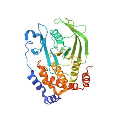
Protein tyrosine phosphatases (PTPs) possess a conserved mobile catalytic loop, the WPD-loop, which brings an aspartic acid into the active site where it acts as an acid/base catalyst. Prior experimental and computational studies, focused on the human enzyme PTP1B and the PTP from Yersinia pestis , YopH, suggested that loop conformational dynamics are important in regulating both catalysis and evolvability. We have generated a chimeric protein in which the WPD-loop of YopH is transposed into PTP1B, and eight chimeras that systematically restored the loop sequence back to native PTP1B. Of these, four chimeras were soluble and were subjected to detailed biochemical and structural characterization, and a computational analysis of their WPD-loop dynamics. The chimeras maintain backbone structural integrity, with somewhat slower rates than either wild-type parent, and show differences in the pH dependency of catalysis, and changes in the effect of Mg 2+ . The chimeric proteins' WPD-loops differ significantly in their relative stability and rigidity. The time required for interconversion, coupled with electrostatic effects revealed by simulations, likely accounts for the activity differences between chimeras, and relative to the native enzymes. Our results further the understanding of connections between enzyme activity and the dynamics of catalytically important groups, particularly the effects of non-catalytic residues on key conformational equilibria.
Organizational Affiliation:
Department of Chemistry and Biochemistry, Utah State University Logan Utah 84322-0300 USA.