6WZZ

GID4 in complex with VGLWKS peptide


Experimental Data Snapshot

  • Method: X-RAY DIFFRACTION
  • Resolution: 1.60 Å
  • R-Value Free: 0.220 
  • R-Value Work: 0.186 

wwPDB Validation   3D Report Full Report


This is version 1.2 of the entry. See complete history


Literature

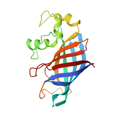
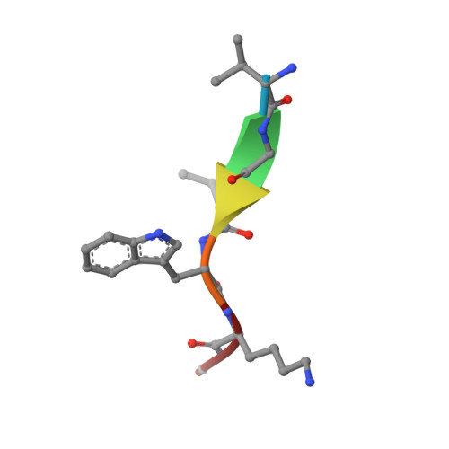
Recognition of nonproline N-terminal residues by the Pro/N-degron pathway.

Dong, C.Chen, S.J.Melnykov, A.Weirich, S.Sun, K.Jeltsch, A.Varshavsky, A.Min, J.

(2020) Proc Natl Acad Sci U S A 117: 14158-14167

  • DOI: https://doi.org/10.1073/pnas.2007085117
  • Primary Citation of Related Structures:  
    6WZX, 6WZZ

  • PubMed Abstract: 

    Eukaryotic N-degron pathways are proteolytic systems whose unifying feature is their ability to recognize proteins containing N-terminal (Nt) degradation signals called N-degrons, and to target these proteins for degradation by the 26S proteasome or autophagy. GID4, a subunit of the GID ubiquitin ligase, is the main recognition component of the proline (Pro)/N-degron pathway. GID4 targets proteins through their Nt-Pro residue or a Pro at position 2, in the presence of specific downstream sequence motifs. Here we show that human GID4 can also recognize hydrophobic Nt-residues other than Pro. One example is the sequence Nt-IGLW, bearing Nt-Ile. Nt-IGLW binds to wild-type human GID4 with a K d of 16 μM, whereas the otherwise identical Nt-Pro-bearing sequence PGLW binds to GID4 more tightly, with a K d of 1.9 μM. Despite this difference in affinities of GID4 for Nt-IGLW vs. Nt-PGLW, we found that the GID4-mediated Pro/N-degron pathway of the yeast Saccharomyces cerevisiae can target an Nt-IGLW-bearing protein for rapid degradation. We solved crystal structures of human GID4 bound to a peptide bearing Nt-Ile or Nt-Val. We also altered specific residues of human GID4 and measured the affinities of resulting mutant GID4s for Nt-IGLW and Nt-PGLW, thereby determining relative contributions of specific GID4 residues to the GID4-mediated recognition of Nt-Pro vs. Nt-residues other than Pro. These and related results advance the understanding of targeting by the Pro/N-degron pathway and greatly expand the substrate recognition range of the GID ubiquitin ligase in both human and yeast cells.


  • Organizational Affiliation

    Department of Biochemistry and Molecular Biology, School of Basic Medical Sciences, Tianjin Medical University, 300070 Tianjin, People's Republic of China.


Macromolecules
Find similar proteins by:  (by identity cutoff)  |  3D Structure
Entity ID: 1
MoleculeChains Sequence LengthOrganismDetailsImage
Glucose-induced degradation protein 4 homolog167Homo sapiensMutation(s): 0 
Gene Names: GID4C17orf39VID24
UniProt & NIH Common Fund Data Resources
Find proteins for Q8IVV7 (Homo sapiens)
Explore Q8IVV7 
Go to UniProtKB:  Q8IVV7
PHAROS:  Q8IVV7
GTEx:  ENSG00000141034 
Entity Groups  
Sequence Clusters30% Identity50% Identity70% Identity90% Identity95% Identity100% Identity
UniProt GroupQ8IVV7
Sequence Annotations
Expand
  • Reference Sequence

Find similar proteins by:  Sequence   |   3D Structure  

Entity ID: 2
MoleculeChains Sequence LengthOrganismDetailsImage
VGLWKS peptide6synthetic constructMutation(s): 0 
Sequence Annotations
Expand
  • Reference Sequence
Small Molecules
Ligands 1 Unique
IDChains Name / Formula / InChI Key2D Diagram3D Interactions
UNX
Query on UNX

Download Ideal Coordinates CCD File 
C [auth A]
D [auth A]
E [auth A]
F [auth A]
G [auth A]
C [auth A],
D [auth A],
E [auth A],
F [auth A],
G [auth A],
H [auth A],
I [auth A],
J [auth A],
K [auth A],
L [auth A],
M [auth A],
N [auth A],
O [auth A]
UNKNOWN ATOM OR ION
X
Experimental Data & Validation

Experimental Data

  • Method: X-RAY DIFFRACTION
  • Resolution: 1.60 Å
  • R-Value Free: 0.220 
  • R-Value Work: 0.186 
  • Space Group: P 1 21 1
Unit Cell:
Length ( Å )Angle ( ˚ )
a = 36.781α = 90
b = 40.648β = 98.102
c = 56.319γ = 90
Software Package:
Software NamePurpose
XDSdata reduction
Aimlessdata scaling
REFMACrefinement
PDB_EXTRACTdata extraction
PHASERphasing

Structure Validation

View Full Validation Report


Entry History 

Deposition Data

Revision History  (Full details and data files)

  • Version 1.0: 2020-06-17
    Type: Initial release
  • Version 1.1: 2020-07-08
    Changes: Database references
  • Version 1.2: 2023-10-18
    Changes: Data collection, Database references, Refinement description