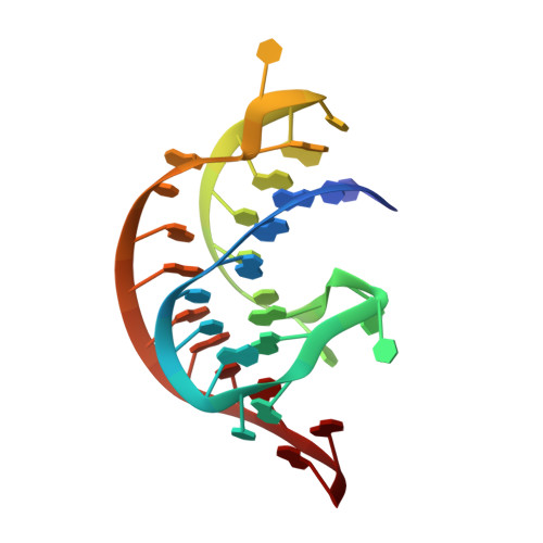
Analysis of a preQ1-I riboswitch in effector-free and bound states reveals a metabolite-programmed nucleobase-stacking spine that controls gene regulation.
Schroeder, G.M., Dutta, D., Cavender, C.E., Jenkins, J.L., Pritchett, E.M., Baker, C.D., Ashton, J.M., Mathews, D.H., Wedekind, J.E.(2020) Nucleic Acids Res 48: 8146-8164
- PubMed: 32597951
- DOI: https://doi.org/10.1093/nar/gkaa546
- Primary Citation of Related Structures:
6VUH, 6VUI - PubMed Abstract:
Riboswitches are structured RNA motifs that recognize metabolites to alter the conformations of downstream sequences, leading to gene regulation. To investigate this molecular framework, we determined crystal structures of a preQ1-I riboswitch in effector-free and bound states at 2.00 Å and 2.65 Å-resolution. Both pseudoknots exhibited the elusive L2 loop, which displayed distinct conformations. Conversely, the Shine-Dalgarno sequence (SDS) in the S2 helix of each structure remained unbroken. The expectation that the effector-free state should expose the SDS prompted us to conduct solution experiments to delineate environmental changes to specific nucleobases in response to preQ1. We then used nudged elastic band computational methods to derive conformational-change pathways linking the crystallographically-determined effector-free and bound-state structures. Pathways featured: (i) unstacking and unpairing of L2 and S2 nucleobases without preQ1-exposing the SDS for translation and (ii) stacking and pairing L2 and S2 nucleobases with preQ1-sequestering the SDS. Our results reveal how preQ1 binding reorganizes L2 into a nucleobase-stacking spine that sequesters the SDS, linking effector recognition to biological function. The generality of stacking spines as conduits for effector-dependent, interdomain communication is discussed in light of their existence in adenine riboswitches, as well as the turnip yellow mosaic virus ribosome sensor.
Organizational Affiliation:
Department of Biochemistry & Biophysics, University of Rochester School of Medicine & Dentistry, Rochester, NY 14642, USA.