6VIN

Crystallographic structure of the circularly permuted human Taspase1 protein


Experimental Data Snapshot

  • Method: X-RAY DIFFRACTION
  • Resolution: 3.04 Å
  • R-Value Free: 0.307 
  • R-Value Work: 0.237 
  • R-Value Observed: 0.243 

wwPDB Validation   3D Report Full Report

Currently 6VIN does not have a validation slider image.


This is version 1.3 of the entry. See complete history


Literature

Structural insights into the function of the catalytically active human Taspase1.

Nagaratnam, N.Delker, S.L.Jernigan, R.Edwards, T.E.Snider, J.Thifault, D.Williams, D.Nannenga, B.L.Stofega, M.Sambucetti, L.Hsieh, J.J.Flint, A.J.Fromme, P.Martin-Garcia, J.M.

(2021) Structure 29: 873

  • DOI: https://doi.org/10.1016/j.str.2021.03.008
  • Primary Citation of Related Structures:  
    6UGK, 6VIN

  • PubMed Abstract: 

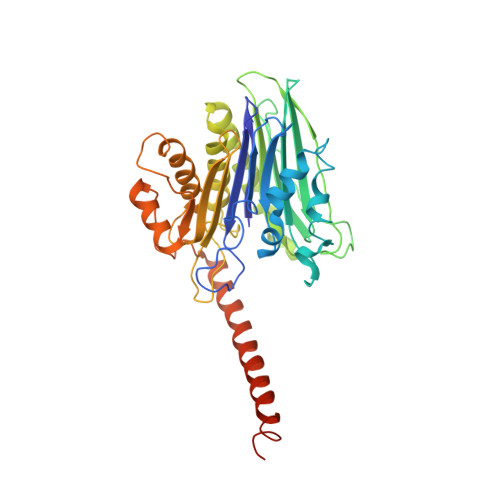
    Taspase1 is an Ntn-hydrolase overexpressed in primary human cancers, coordinating cancer cell proliferation, invasion, and metastasis. Loss of Taspase1 activity disrupts proliferation of human cancer cells in vitro and in mouse models of glioblastoma. Taspase1 is synthesized as an inactive proenzyme, becoming active upon intramolecular cleavage. The activation process changes the conformation of a long fragment at the C-terminus of the α subunit, for which no full-length structural information exists and whose function is poorly understood. We present a cloning strategy to generate a circularly permuted form of Taspase1 to determine the crystallographic structure of active Taspase1. We discovered that this region forms a long helix and is indispensable for the catalytic activity of Taspase1. Our study highlights the importance of this element for the enzymatic activity of Ntn-hydrolases, suggesting that it could be a potential target for the design of inhibitors with potential to be developed into anticancer therapeutics.


  • Organizational Affiliation

    Center for Applied Structural Discovery, Biodesign Institute, Arizona State University, Tempe, AZ 85287, USA.


Macromolecules
Find similar proteins by:  (by identity cutoff)  |  3D Structure
Entity ID: 1
MoleculeChains Sequence LengthOrganismDetailsImage
Threonine aspartase 1
A, B
389Homo sapiensMutation(s): 0 
Gene Names: TASP1C20orf13
EC: 3.4.25
UniProt & NIH Common Fund Data Resources
Find proteins for Q9H6P5 (Homo sapiens)
Explore Q9H6P5 
Go to UniProtKB:  Q9H6P5
PHAROS:  Q9H6P5
GTEx:  ENSG00000089123 
Entity Groups  
Sequence Clusters30% Identity50% Identity70% Identity90% Identity95% Identity100% Identity
UniProt GroupQ9H6P5
Sequence Annotations
Expand
  • Reference Sequence
Experimental Data & Validation

Experimental Data

  • Method: X-RAY DIFFRACTION
  • Resolution: 3.04 Å
  • R-Value Free: 0.307 
  • R-Value Work: 0.237 
  • R-Value Observed: 0.243 
  • Space Group: H 3 2
Unit Cell:
Length ( Å )Angle ( ˚ )
a = 196α = 90
b = 196β = 90
c = 196.91γ = 120
Software Package:
Software NamePurpose
REFMACrefinement
PDB_EXTRACTdata extraction
XDSdata reduction
STARANISOdata scaling
PHASERphasing

Structure Validation

View Full Validation Report

Currently 6VIN does not have a validation slider image.


Entry History & Funding Information

Deposition Data


Funding OrganizationLocationGrant Number
National Institutes of Health/National Cancer Institute (NIH/NCI)United StatesNX118-107-160-007

Revision History  (Full details and data files)

  • Version 1.0: 2021-01-13
    Type: Initial release
  • Version 1.1: 2021-04-14
    Changes: Database references
  • Version 1.2: 2021-08-18
    Changes: Database references
  • Version 1.3: 2023-10-11
    Changes: Data collection, Refinement description