6VGD

Crystal structure of the DNA binding domain (DBD) of human FLI1 and the complex of the DBD of human Runx2 with core binding factor beta (Cbfb), in complex with 16mer DNA CAGAGGATGTGGCTTC


Experimental Data Snapshot

  • Method: X-RAY DIFFRACTION
  • Resolution: 4.20 Å
  • R-Value Free: 0.293 
  • R-Value Work: 0.295 
  • R-Value Observed: 0.295 

wwPDB Validation   3D Report Full Report

Currently 6VGD does not have a validation slider image.


This is version 1.3 of the entry. See complete history


Literature

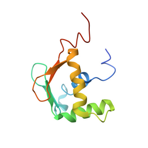
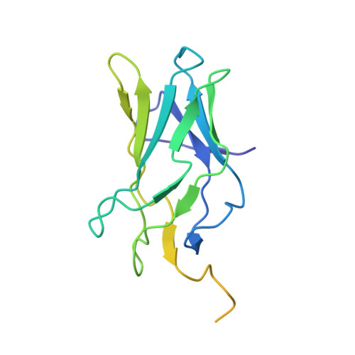
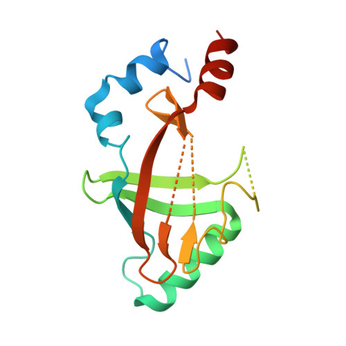
Allosteric interference in oncogenic FLI1 and ERG transactions by mithramycins.

Hou, C.Mandal, A.Rohr, J.Tsodikov, O.V.

(2021) Structure 29: 404-412.e4

  • DOI: https://doi.org/10.1016/j.str.2020.11.012
  • Primary Citation of Related Structures:  
    6VG2, 6VG8, 6VGD, 6VGE, 6VGG

  • PubMed Abstract: 

    ETS family transcription factors of ERG and FLI1 play a key role in oncogenesis of prostate cancer and Ewing sarcoma by binding regulatory DNA sites and interfering with function of other factors. Mithramycin (MTM) is an anti-cancer, DNA binding natural product that functions as a potent antagonist of ERG and FLI1 by an unknown mechanism. We present a series of crystal structures of the DNA binding domain (DBD) of ERG/FLI1 culminating in a structure of a high-order complex of the ERG/FLI1 DBD, transcription factor Runx2, core-binding factor beta (Cbfβ), and MTM on a DNA enhancer site, along with supporting DNA binding studies using MTM and its analogues. Taken together, these data provide insight into allosteric mechanisms underlying ERG and FLI1 transactions and their disruption by MTM analogues.


  • Organizational Affiliation

    Department of Pharmaceutical Sciences, College of Pharmacy, University of Kentucky, Lexington, KY 40536, USA.


Macromolecules

Find similar proteins by:  (by identity cutoff)  |  3D Structure
Entity ID: 1
MoleculeChains Sequence LengthOrganismDetailsImage
Friend leukemia integration 1 transcription factor104Homo sapiensMutation(s): 0 
Gene Names: FLI1
UniProt & NIH Common Fund Data Resources
Find proteins for Q01543 (Homo sapiens)
Explore Q01543 
Go to UniProtKB:  Q01543
PHAROS:  Q01543
GTEx:  ENSG00000151702 
Entity Groups  
Sequence Clusters30% Identity50% Identity70% Identity90% Identity95% Identity100% Identity
UniProt GroupQ01543
Sequence Annotations
Expand
  • Reference Sequence
Find similar proteins by:  (by identity cutoff)  |  3D Structure
Entity ID: 4
MoleculeChains Sequence LengthOrganismDetailsImage
Runt-related transcription factor 2177Homo sapiensMutation(s): 0 
Gene Names: RUNX2AML3CBFA1OSF2PEBP2A
UniProt & NIH Common Fund Data Resources
Find proteins for Q13950 (Homo sapiens)
Explore Q13950 
Go to UniProtKB:  Q13950
PHAROS:  Q13950
GTEx:  ENSG00000124813 
Entity Groups  
Sequence Clusters30% Identity50% Identity70% Identity90% Identity95% Identity100% Identity
UniProt GroupQ13950
Sequence Annotations
Expand
  • Reference Sequence
Find similar proteins by:  (by identity cutoff)  |  3D Structure
Entity ID: 5
MoleculeChains Sequence LengthOrganismDetailsImage
Core-binding factor subunit betaE [auth G]156Homo sapiensMutation(s): 0 
Gene Names: CBFB
UniProt & NIH Common Fund Data Resources
Find proteins for Q13951 (Homo sapiens)
Explore Q13951 
Go to UniProtKB:  Q13951
PHAROS:  Q13951
GTEx:  ENSG00000067955 
Entity Groups  
Sequence Clusters30% Identity50% Identity70% Identity90% Identity95% Identity100% Identity
UniProt GroupQ13951
Sequence Annotations
Expand
  • Reference Sequence

Find similar nucleic acids by:  Sequence   |   3D Structure  

Entity ID: 2
MoleculeChains LengthOrganismImage
DNA (5'-D(P*CP*AP*GP*AP*GP*GP*AP*TP*GP*TP*GP*GP*CP*TP*TP*C)-3')16Homo sapiens
Sequence Annotations
Expand
  • Reference Sequence

Find similar nucleic acids by:  Sequence   |   3D Structure  

Entity ID: 3
MoleculeChains LengthOrganismImage
DNA (5'-D(P*GP*AP*AP*GP*CP*CP*AP*CP*AP*TP*CP*CP*TP*CP*TP*G)-3')16Homo sapiens
Sequence Annotations
Expand
  • Reference Sequence
Experimental Data & Validation

Experimental Data

  • Method: X-RAY DIFFRACTION
  • Resolution: 4.20 Å
  • R-Value Free: 0.293 
  • R-Value Work: 0.295 
  • R-Value Observed: 0.295 
  • Space Group: P 62 2 2
Unit Cell:
Length ( Å )Angle ( ˚ )
a = 104.513α = 90
b = 104.513β = 90
c = 322.04γ = 120
Software Package:
Software NamePurpose
REFMACrefinement
PDB_EXTRACTdata extraction
HKL-2000data reduction
HKL-2000data scaling
PHASERphasing

Structure Validation

View Full Validation Report

Currently 6VGD does not have a validation slider image.


Entry History & Funding Information

Deposition Data


Funding OrganizationLocationGrant Number
Department of Defense (DOD, United States)United StatesPC150300P1
Department of Defense (DOD, United States)United StatesPC150300P2

Revision History  (Full details and data files)

  • Version 1.0: 2020-11-25
    Type: Initial release
  • Version 1.1: 2020-12-16
    Changes: Database references
  • Version 1.2: 2021-05-19
    Changes: Database references
  • Version 1.3: 2023-10-11
    Changes: Data collection, Database references, Refinement description