


1.6 Angstrom Resolution Crystal Structure of endoglucanase from Komagataeibacter sucrofermentans
Wu, R., Kim, Y., Jedrzejczak, R., Joachimiak, A.To be published.
Experimental Data Snapshot
wwPDB Validation 3D Report Full Report
Currently 6VC5 does not have a validation slider image.
Entity ID: 1 | |||||
|---|---|---|---|---|---|
| Molecule | Chains | Sequence Length | Organism | Details | Image |
| Endoglucanase | 321 | Komagataeibacter sucrofermentans | Mutation(s): 0 Gene Names: CMCase | 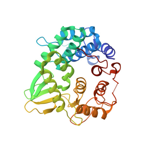 | |
UniProt | |||||
Find proteins for O82857 (Komagataeibacter sucrofermentans) Explore O82857 Go to UniProtKB: O82857 | |||||
Entity Groups | |||||
| Sequence Clusters | 30% Identity50% Identity70% Identity90% Identity95% Identity100% Identity | ||||
| UniProt Group | O82857 | ||||
Sequence AnnotationsExpand | |||||
| |||||
| Ligands 1 Unique | |||||
|---|---|---|---|---|---|
| ID | Chains | Name / Formula / InChI Key | 2D Diagram | 3D Interactions | |
| GOL Query on GOL | B [auth A], C [auth A], D [auth A], E [auth A] | GLYCEROL C3 H8 O3 PEDCQBHIVMGVHV-UHFFFAOYSA-N |  | ||
| Length ( Å ) | Angle ( ˚ ) |
|---|---|
| a = 120.52 | α = 90 |
| b = 120.52 | β = 90 |
| c = 47.588 | γ = 90 |
| Software Name | Purpose |
|---|---|
| PHENIX | refinement |
| SCALEPACK | data scaling |
| PDB_EXTRACT | data extraction |
| HKL-3000 | data reduction |
| HKL-3000 | phasing |
Currently 6VC5 does not have a validation slider image.
RCSB PDB (citation) is hosted by
RCSB PDB is a member of the

