6V86

Parainfluenza virus 5 L-P complex with an alternate conformation of the CD-MTase-CTD module

  • Classification: VIRAL PROTEIN
  • Organism(s): Simian virus 5 (strain W3)
  • Expression System: Spodoptera frugiperda
  • Mutation(s): No 

  • Deposited: 2019-12-10 Released: 2020-02-19 
  • Deposition Author(s): Abdella, R., He, Y.
  • Funding Organization(s): Howard Hughes Medical Institute (HHMI), National Institutes of Health/National Cancer Institute (NIH/NCI), American Cancer Society, National Institutes of Health/National Institute of General Medical Sciences (NIH/NIGMS)

Experimental Data Snapshot

  • Method: ELECTRON MICROSCOPY
  • Resolution: 4.63 Å
  • Aggregation State: PARTICLE 
  • Reconstruction Method: SINGLE PARTICLE 

wwPDB Validation   3D Report Full Report


This is version 1.3 of the entry. See complete history


Literature

Structure of a paramyxovirus polymerase complex reveals a unique methyltransferase-CTD conformation.

Abdella, R.Aggarwal, M.Okura, T.Lamb, R.A.He, Y.

(2020) Proc Natl Acad Sci U S A 117: 4931-4941

  • DOI: https://doi.org/10.1073/pnas.1919837117
  • Primary Citation of Related Structures:  
    6V85, 6V86, 6VAG

  • PubMed Abstract: 

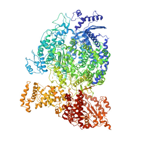
    Paramyxoviruses are enveloped, nonsegmented, negative-strand RNA viruses that cause a wide spectrum of human and animal diseases. The viral genome, packaged by the nucleoprotein (N), serves as a template for the polymerase complex, composed of the large protein (L) and the homo-tetrameric phosphoprotein (P). The ∼250-kDa L possesses all enzymatic activities necessary for its function but requires P in vivo. Structural information is available for individual P domains from different paramyxoviruses, but how P interacts with L and how that affects the activity of L is largely unknown due to the lack of high-resolution structures of this complex in this viral family. In this study we determined the structure of the L-P complex from parainfluenza virus 5 (PIV5) at 4.3-Å resolution using cryoelectron microscopy, as well as the oligomerization domain (OD) of P at 1.4-Å resolution using X-ray crystallography. P-OD associates with the RNA-dependent RNA polymerase domain of L and protrudes away from it, while the X domain of one chain of P is bound near the L nucleotide entry site. The methyltransferase (MTase) domain and the C-terminal domain (CTD) of L adopt a unique conformation, positioning the MTase active site immediately above the poly-ribonucleotidyltransferase domain and near the likely exit site for the product RNA 5' end. Our study reveals a potential mechanism that mononegavirus polymerases may employ to switch between transcription and genome replication. This knowledge will assist in the design and development of antivirals against paramyxoviruses.


  • Organizational Affiliation

    Department of Molecular Biosciences, Northwestern University, Evanston, IL 60208.


Macromolecules
Find similar proteins by:  (by identity cutoff)  |  3D Structure
Entity ID: 1
MoleculeChains Sequence LengthOrganismDetailsImage
RNA-directed RNA polymerase L2,255Simian virus 5 (strain W3)Mutation(s): 0 
EC: 2.7.7.48 (PDB Primary Data), 2.1.1.56 (PDB Primary Data), 2.7.7.88 (PDB Primary Data), 2.1.1.296 (PDB Primary Data)
UniProt
Find proteins for Q88434 (Parainfluenza virus 5 (strain W3))
Explore Q88434 
Go to UniProtKB:  Q88434
Entity Groups  
Sequence Clusters30% Identity50% Identity70% Identity90% Identity95% Identity100% Identity
UniProt GroupQ88434
Sequence Annotations
Expand
  • Reference Sequence
Find similar proteins by:  (by identity cutoff)  |  3D Structure
Entity ID: 2
MoleculeChains Sequence LengthOrganismDetailsImage
Phosphoprotein
B, C, D, E, F
392Simian virus 5 (strain W3)Mutation(s): 0 
Gene Names: P/V
UniProt
Find proteins for P11208 (Parainfluenza virus 5 (strain W3))
Explore P11208 
Go to UniProtKB:  P11208
Entity Groups  
Sequence Clusters30% Identity50% Identity70% Identity90% Identity95% Identity100% Identity
UniProt GroupP11208
Sequence Annotations
Expand
  • Reference Sequence
Experimental Data & Validation

Experimental Data

  • Method: ELECTRON MICROSCOPY
  • Resolution: 4.63 Å
  • Aggregation State: PARTICLE 
  • Reconstruction Method: SINGLE PARTICLE 
EM Software:
TaskSoftware PackageVersion
RECONSTRUCTIONRELION3.1

Structure Validation

View Full Validation Report


Entry History & Funding Information

Deposition Data


Funding OrganizationLocationGrant Number
Howard Hughes Medical Institute (HHMI)United States--
National Institutes of Health/National Cancer Institute (NIH/NCI)United StatesU54-CA193419
National Institutes of Health/National Cancer Institute (NIH/NCI)United StatesP01-CA092584
American Cancer SocietyUnited StatesIRG-15-173-21
National Institutes of Health/National Institute of General Medical Sciences (NIH/NIGMS)United StatesT32-GM008382

Revision History  (Full details and data files)

  • Version 1.0: 2020-02-19
    Type: Initial release
  • Version 1.1: 2020-03-04
    Changes: Database references
  • Version 1.2: 2020-03-18
    Changes: Database references
  • Version 1.3: 2024-03-06
    Changes: Data collection, Database references