6UZ1

Noncanonical binding of single-chain A6 TCR variant S3-4 in complex with Tax/HLA-A2


Experimental Data Snapshot

  • Method: X-RAY DIFFRACTION
  • Resolution: 3.14 Å
  • R-Value Free: 0.239 
  • R-Value Work: 0.189 
  • R-Value Observed: 0.192 

wwPDB Validation   3D Report Full Report

Currently 6UZ1 does not have a validation slider image.


This is version 1.2 of the entry. See complete history


Literature

An Engineered T Cell Receptor Variant Realizes the Limits of Functional Binding Modes.

Singh, N.K.Alonso, J.A.Harris, D.T.Anderson, S.D.Ma, J.Hellman, L.M.Rosenberg, A.M.Kolawole, E.M.Evavold, B.D.Kranz, D.M.Baker, B.M.

(2020) Biochemistry 59: 4163-4175

  • DOI: https://doi.org/10.1021/acs.biochem.0c00689
  • Primary Citation of Related Structures:  
    6UZ1

  • PubMed Abstract: 

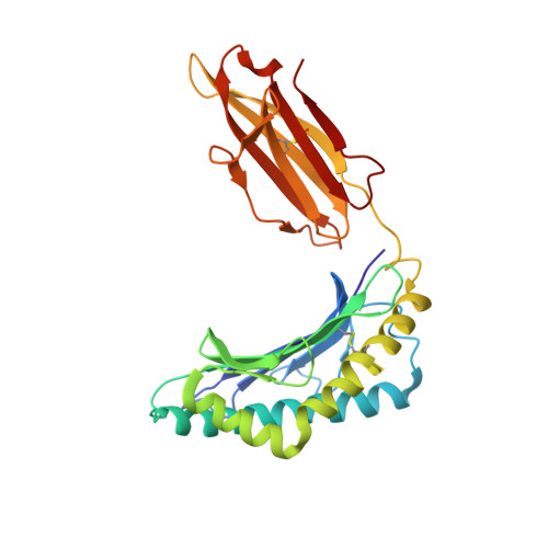
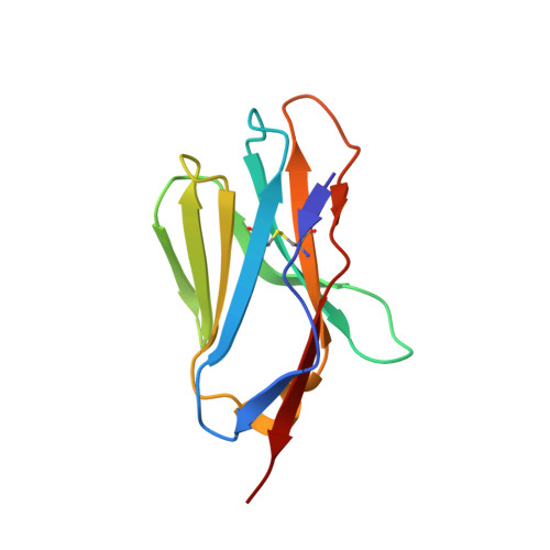
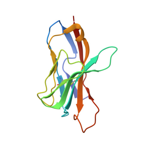
    T cell receptors (TCRs) orchestrate cellular immunity by recognizing peptides presented by a range of major histocompatibility complex (MHC) proteins. Naturally occurring TCRs bind the composite peptide/MHC surface, recognizing peptides that are structurally and chemically compatible with the TCR binding site. Here we describe a molecularly evolved TCR variant that binds the human class I MHC protein HLA-A2 independent of the bound peptide, achieved by a drastic perturbation of the TCR binding geometry that places the molecule far from the peptide binding groove. This unique geometry is unsupportive of normal T cell signaling. A substantial divergence between affinity measurements in solution and in two dimensions between proximal cell membranes leads us to attribute the lack of signaling to steric hindrance that limits binding in the confines of a cell-cell interface. Our results provide an example of how receptor binding geometry can impact T cell function and provide further support for the view that germline-encoded residues in TCR binding loops evolved to drive productive TCR recognition and signaling.


  • Organizational Affiliation

    Department of Chemistry and Biochemistry and Harper Cancer Research Institute, University of Notre Dame, Notre Dame, Indiana 46556, United States.


Macromolecules
Find similar proteins by:  (by identity cutoff)  |  3D Structure
Entity ID: 1
MoleculeChains Sequence LengthOrganismDetailsImage
MHC class I antigen, A-2 alpha chainA,
D [auth F]
275Homo sapiensMutation(s): 0 
Gene Names: HLA-A
UniProt
Find proteins for A0A140T913 (Homo sapiens)
Explore A0A140T913 
Go to UniProtKB:  A0A140T913
Entity Groups  
Sequence Clusters30% Identity50% Identity70% Identity90% Identity95% Identity100% Identity
UniProt GroupA0A140T913
Sequence Annotations
Expand
  • Reference Sequence
Find similar proteins by:  (by identity cutoff)  |  3D Structure
Entity ID: 2
MoleculeChains Sequence LengthOrganismDetailsImage
Beta-2-microglobulinB,
E [auth G]
100Homo sapiensMutation(s): 0 
Gene Names: B2MCDABP0092HDCMA22P
UniProt & NIH Common Fund Data Resources
Find proteins for P61769 (Homo sapiens)
Explore P61769 
Go to UniProtKB:  P61769
PHAROS:  P61769
GTEx:  ENSG00000166710 
Entity Groups  
Sequence Clusters30% Identity50% Identity70% Identity90% Identity95% Identity100% Identity
UniProt GroupP61769
Sequence Annotations
Expand
  • Reference Sequence

Find similar proteins by:  Sequence   |   3D Structure  

Entity ID: 3
MoleculeChains Sequence LengthOrganismDetailsImage
LEU-LEU-PHE-GLY-TYR-PRO-VAL-TYR-VALC,
F [auth H]
9Homo sapiensMutation(s): 0 
UniProt
Find proteins for P14079 (Human T-cell leukemia virus 1 (isolate Caribbea HS-35 subtype A))
Explore P14079 
Go to UniProtKB:  P14079
Entity Groups  
UniProt GroupP14079
Sequence Annotations
Expand
  • Reference Sequence
Find similar proteins by:  (by identity cutoff)  |  3D Structure
Entity ID: 4
MoleculeChains Sequence LengthOrganismDetailsImage
T cell receptor, alpha chainG [auth I],
I [auth D]
110Homo sapiensMutation(s): 0 
Entity Groups  
Sequence Clusters30% Identity50% Identity70% Identity90% Identity95% Identity100% Identity
Sequence Annotations
Expand
  • Reference Sequence
Find similar proteins by:  (by identity cutoff)  |  3D Structure
Entity ID: 5
MoleculeChains Sequence LengthOrganismDetailsImage
T cell receptor, beta chainH [auth J],
J [auth E]
115Homo sapiensMutation(s): 0 
Entity Groups  
Sequence Clusters30% Identity50% Identity70% Identity90% Identity95% Identity100% Identity
Sequence Annotations
Expand
  • Reference Sequence
Small Molecules
Ligands 1 Unique
IDChains Name / Formula / InChI Key2D Diagram3D Interactions
MES
Query on MES

Download Ideal Coordinates CCD File 
K [auth B],
L [auth F]
2-(N-MORPHOLINO)-ETHANESULFONIC ACID
C6 H13 N O4 S
SXGZJKUKBWWHRA-UHFFFAOYSA-N
Experimental Data & Validation

Experimental Data

  • Method: X-RAY DIFFRACTION
  • Resolution: 3.14 Å
  • R-Value Free: 0.239 
  • R-Value Work: 0.189 
  • R-Value Observed: 0.192 
  • Space Group: P 61
Unit Cell:
Length ( Å )Angle ( ˚ )
a = 236.65α = 90
b = 236.65β = 90
c = 63.146γ = 120
Software Package:
Software NamePurpose
PHENIXrefinement
PHENIXrefinement
HKL-2000data reduction
HKL-2000data scaling
PHENIXphasing

Structure Validation

View Full Validation Report

Currently 6UZ1 does not have a validation slider image.


Entry History 

Deposition Data

Revision History  (Full details and data files)

  • Version 1.0: 2020-10-28
    Type: Initial release
  • Version 1.1: 2020-11-11
    Changes: Database references
  • Version 1.2: 2023-10-11
    Changes: Data collection, Database references, Refinement description