6UOA

Human metabotropic GABA(B) receptor in its intermediate state 1


Experimental Data Snapshot

  • Method: ELECTRON MICROSCOPY
  • Resolution: 6.30 Å
  • Aggregation State: PARTICLE 
  • Reconstruction Method: SINGLE PARTICLE 

wwPDB Validation   3D Report Full Report


This is version 2.1 of the entry. See complete history


Literature

Structural basis of the activation of a metabotropic GABA receptor.

Shaye, H.Ishchenko, A.Lam, J.H.Han, G.W.Xue, L.Rondard, P.Pin, J.P.Katritch, V.Gati, C.Cherezov, V.

(2020) Nature 584: 298-303

  • DOI: https://doi.org/10.1038/s41586-020-2408-4
  • Primary Citation of Related Structures:  
    6UO8, 6UO9, 6UOA, 6VJM

  • PubMed Abstract: 

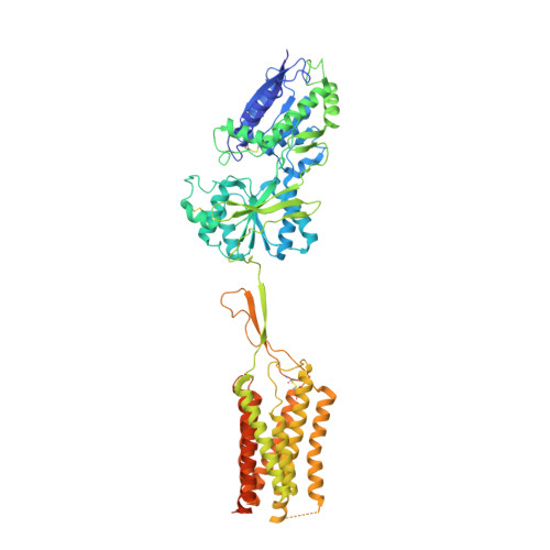
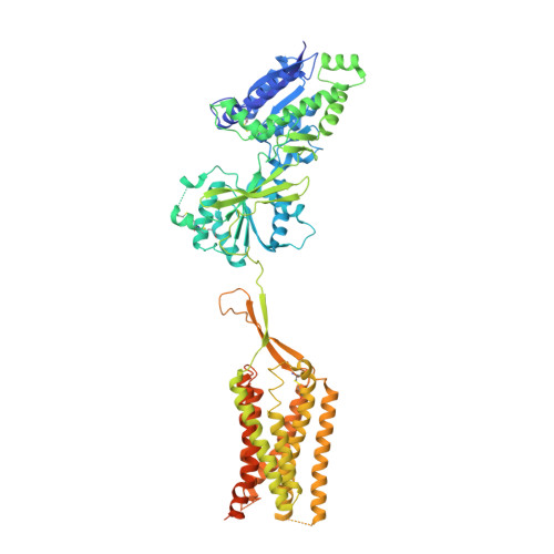
    Metabotropic γ-aminobutyric acid receptors (GABA B ) are involved in the modulation of synaptic responses in the central nervous system and have been implicated in neuropsychological conditions that range from addiction to psychosis 1 . GABA B belongs to class C of the G-protein-coupled receptors, and its functional entity comprises an obligate heterodimer that is composed of the GB1 and GB2 subunits 2 . Each subunit possesses an extracellular Venus flytrap domain, which is connected to a canonical seven-transmembrane domain. Here we present four cryo-electron microscopy structures of the human full-length GB1-GB2 heterodimer: one structure of its inactive apo state, two intermediate agonist-bound forms and an active form in which the heterodimer is bound to an agonist and a positive allosteric modulator. The structures reveal substantial differences, which shed light on the complex motions that underlie the unique activation mechanism of GABA B . Our results show that agonist binding leads to the closure of the Venus flytrap domain of GB1, triggering a series of transitions, first rearranging and bringing the two transmembrane domains into close contact along transmembrane helix 6 and ultimately inducing conformational rearrangements in the GB2 transmembrane domain via a lever-like mechanism to initiate downstream signalling. This active state is stabilized by a positive allosteric modulator binding at the transmembrane dimerization interface.


  • Organizational Affiliation

    Bridge Institute, USC Michelson Center for Convergent Biosciences, University of Southern California, Los Angeles, CA, USA.


Macromolecules
Find similar proteins by:  (by identity cutoff)  |  3D Structure
Entity ID: 1
MoleculeChains Sequence LengthOrganismDetailsImage
Gamma-aminobutyric acid type B receptor subunit 1762Homo sapiensMutation(s): 0 
Gene Names: GABBR1GPRC3A
Membrane Entity: Yes 
UniProt & NIH Common Fund Data Resources
Find proteins for Q9UBS5 (Homo sapiens)
Explore Q9UBS5 
Go to UniProtKB:  Q9UBS5
PHAROS:  Q9UBS5
GTEx:  ENSG00000204681 
Entity Groups  
Sequence Clusters30% Identity50% Identity70% Identity90% Identity95% Identity100% Identity
UniProt GroupQ9UBS5
Sequence Annotations
Expand
  • Reference Sequence
Find similar proteins by:  (by identity cutoff)  |  3D Structure
Entity ID: 2
MoleculeChains Sequence LengthOrganismDetailsImage
Gamma-aminobutyric acid type B receptor subunit 2779Homo sapiensMutation(s): 0 
Gene Names: GABBR2GPR51GPRC3B
Membrane Entity: Yes 
UniProt & NIH Common Fund Data Resources
Find proteins for O75899 (Homo sapiens)
Explore O75899 
Go to UniProtKB:  O75899
PHAROS:  O75899
GTEx:  ENSG00000136928 
Entity Groups  
Sequence Clusters30% Identity50% Identity70% Identity90% Identity95% Identity100% Identity
UniProt GroupO75899
Sequence Annotations
Expand
  • Reference Sequence
Oligosaccharides

Help

Entity ID: 3
MoleculeChains Length2D Diagram Glycosylation3D Interactions
2-acetamido-2-deoxy-beta-D-glucopyranose-(1-4)-2-acetamido-2-deoxy-beta-D-glucopyranose
C, D, E, F, G
2N-Glycosylation
Glycosylation Resources
GlyTouCan:  G42666HT
GlyCosmos:  G42666HT
GlyGen:  G42666HT
Small Molecules
Ligands 1 Unique
IDChains Name / Formula / InChI Key2D Diagram3D Interactions
NAG
Query on NAG

Download Ideal Coordinates CCD File 
H [auth B]2-acetamido-2-deoxy-beta-D-glucopyranose
C8 H15 N O6
OVRNDRQMDRJTHS-FMDGEEDCSA-N
Experimental Data & Validation

Experimental Data

  • Method: ELECTRON MICROSCOPY
  • Resolution: 6.30 Å
  • Aggregation State: PARTICLE 
  • Reconstruction Method: SINGLE PARTICLE 
EM Software:
TaskSoftware PackageVersion
MODEL REFINEMENTPHENIX1.17.1-3660
RECONSTRUCTIONcryoSPARC2.12.14

Structure Validation

View Full Validation Report


Entry History & Funding Information

Deposition Data


Funding OrganizationLocationGrant Number
National Institutes of Health/National Institute of General Medical Sciences (NIH/NIGMS)United StatesGM127086
Department of Energy (DOE, United States)United StatesDE-AC02-76SF00515

Revision History  (Full details and data files)

  • Version 1.0: 2020-06-10
    Type: Initial release
  • Version 1.1: 2020-07-01
    Changes: Database references
  • Version 2.0: 2020-07-29
    Type: Remediation
    Reason: Carbohydrate remediation
    Changes: Advisory, Atomic model, Data collection, Derived calculations, Structure summary
  • Version 2.1: 2020-08-26
    Changes: Database references, Structure summary