Structural basis of peptidoglycan endopeptidase regulation.
Shin, J.H., Sulpizio, A.G., Kelley, A., Alvarez, L., Murphy, S.G., Fan, L., Cava, F., Mao, Y., Saper, M.A., Dorr, T.(2020) Proc Natl Acad Sci U S A 117: 11692-11702
- PubMed: 32393643
- DOI: https://doi.org/10.1073/pnas.2001661117
- Primary Citation of Related Structures:
6U2A, 6UE4 - PubMed Abstract:
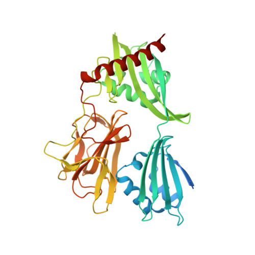
Most bacteria surround themselves with a cell wall, a strong meshwork consisting primarily of the polymerized aminosugar peptidoglycan (PG). PG is essential for structural maintenance of bacterial cells, and thus for viability. PG is also constantly synthesized and turned over; the latter process is mediated by PG cleavage enzymes, for example, the endopeptidases (EPs). EPs themselves are essential for growth but also promote lethal cell wall degradation after exposure to antibiotics that inhibit PG synthases (e.g., β-lactams). Thus, EPs are attractive targets for novel antibiotics and their adjuvants. However, we have a poor understanding of how these enzymes are regulated in vivo, depriving us of novel pathways for the development of such antibiotics. Here, we have solved crystal structures of the LysM/M23 family peptidase ShyA, the primary EP of the cholera pathogen Vibrio cholerae Our data suggest that ShyA assumes two drastically different conformations: a more open form that allows for substrate binding and a closed form, which we predicted to be catalytically inactive. Mutations expected to promote the open conformation caused enhanced activity in vitro and in vivo, and these results were recapitulated in EPs from the divergent pathogens Neisseria gonorrheae and Escherichia coli Our results suggest that LysM/M23 EPs are regulated via release of the inhibitory Domain 1 from the M23 active site, likely through conformational rearrangement in vivo.
Organizational Affiliation:
Weill Institute for Cell and Molecular Biology, Cornell University, Ithaca, NY 14853.