6U2H

BRAF dimer bound to 14-3-3


Experimental Data Snapshot

  • Method: X-RAY DIFFRACTION
  • Resolution: 2.50 Å
  • R-Value Free: 0.263 
  • R-Value Work: 0.220 
  • R-Value Observed: 0.222 

wwPDB Validation   3D Report Full Report


Ligand Structure Quality Assessment 


This is version 1.3 of the entry. See complete history


Literature

Negative regulation of RAF kinase activity by ATP is overcome by 14-3-3-induced dimerization.

Liau, N.P.D.Wendorff, T.J.Quinn, J.G.Steffek, M.Phung, W.Liu, P.Tang, J.Irudayanathan, F.J.Izadi, S.Shaw, A.S.Malek, S.Hymowitz, S.G.Sudhamsu, J.

(2020) Nat Struct Mol Biol 27: 134-141

  • DOI: https://doi.org/10.1038/s41594-019-0365-0
  • Primary Citation of Related Structures:  
    6U2G, 6U2H

  • PubMed Abstract: 

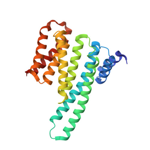
    The RAS-RAF-MEK-ERK signaling axis is frequently activated in human cancers. Physiological concentrations of ATP prevent formation of RAF kinase-domain (RAF KD ) dimers that are critical for activity. Here we present a 2.9-Å-resolution crystal structure of human BRAF KD in complex with MEK and the ATP analog AMP-PCP, revealing interactions between BRAF and ATP that induce an inactive, monomeric conformation of BRAF KD . We also determine how 14-3-3 relieves the negative regulatory effect of ATP through a 2.5-Å-resolution crystal structure of the BRAF KD -14-3-3 complex, in which dimeric 14-3-3 enforces a dimeric BRAF KD assembly to increase BRAF activity. Our data suggest that most oncogenic BRAF mutations alter interactions with ATP and counteract the negative effects of ATP binding by lowering the threshold for RAF dimerization and pathway activation. Our study establishes a framework for rationalizing oncogenic BRAF mutations and provides new avenues for improved RAF-inhibitor discovery.


  • Organizational Affiliation

    Department of Structural Biology, Genentech Inc., South San Francisco, USA.


Macromolecules
Find similar proteins by:  (by identity cutoff)  |  3D Structure
Entity ID: 1
MoleculeChains Sequence LengthOrganismDetailsImage
14-3-3 protein zeta/delta
A, B
232Homo sapiensMutation(s): 0 
Gene Names: YWHAZ
UniProt & NIH Common Fund Data Resources
Find proteins for P63104 (Homo sapiens)
Explore P63104 
Go to UniProtKB:  P63104
PHAROS:  P63104
GTEx:  ENSG00000164924 
Entity Groups  
Sequence Clusters30% Identity50% Identity70% Identity90% Identity95% Identity100% Identity
UniProt GroupP63104
Sequence Annotations
Expand
  • Reference Sequence
Find similar proteins by:  (by identity cutoff)  |  3D Structure
Entity ID: 2
MoleculeChains Sequence LengthOrganismDetailsImage
Serine/threonine-protein kinase B-raf
C, D
290Homo sapiensMutation(s): 0 
Gene Names: BRAFBRAF1RAFB1
EC: 2.7.11.1
UniProt & NIH Common Fund Data Resources
Find proteins for P15056 (Homo sapiens)
Explore P15056 
Go to UniProtKB:  P15056
PHAROS:  P15056
GTEx:  ENSG00000157764 
Entity Groups  
Sequence Clusters30% Identity50% Identity70% Identity90% Identity95% Identity100% Identity
UniProt GroupP15056
Sequence Annotations
Expand
  • Reference Sequence
Small Molecules
Ligands 1 Unique
IDChains Name / Formula / InChI Key2D Diagram3D Interactions
29L
Query on 29L

Download Ideal Coordinates CCD File 
E [auth C],
F [auth D]
2-{4-[(1E)-1-(hydroxyimino)-2,3-dihydro-1H-inden-5-yl]-3-(pyridin-4-yl)-1H-pyrazol-1-yl}ethanol
C19 H18 N4 O2
DEZZLWQELQORIU-RELWKKBWSA-N
Modified Residues  1 Unique
IDChains TypeFormula2D DiagramParent
SEP
Query on SEP
C, D
L-PEPTIDE LINKINGC3 H8 N O6 PSER
Binding Affinity Annotations 
IDSourceBinding Affinity
29L BindingDB:  6U2H Ki: 0.13 (nM) from 1 assay(s)
IC50: min: 0.17, max: 1000 (nM) from 3 assay(s)
Experimental Data & Validation

Experimental Data

  • Method: X-RAY DIFFRACTION
  • Resolution: 2.50 Å
  • R-Value Free: 0.263 
  • R-Value Work: 0.220 
  • R-Value Observed: 0.222 
  • Space Group: P 21 21 21
Unit Cell:
Length ( Å )Angle ( ˚ )
a = 61.504α = 90
b = 115.752β = 90
c = 153.092γ = 90
Software Package:
Software NamePurpose
PHENIXrefinement
PDB_EXTRACTdata extraction
XDSdata reduction
XDSdata scaling
PHENIXphasing

Structure Validation

View Full Validation Report


Ligand Structure Quality Assessment 


Entry History 

Deposition Data

Revision History  (Full details and data files)

  • Version 1.0: 2019-08-28
    Type: Initial release
  • Version 1.1: 2020-02-05
    Changes: Database references
  • Version 1.2: 2020-02-26
    Changes: Database references
  • Version 1.3: 2023-10-11
    Changes: Data collection, Database references, Refinement description