6TQ4

Crystal structure of the Orexin-1 receptor in complex with Compound 16


Experimental Data Snapshot

  • Method: X-RAY DIFFRACTION
  • Resolution: 2.30 Å
  • R-Value Free: 0.236 
  • R-Value Work: 0.209 
  • R-Value Observed: 0.210 

wwPDB Validation   3D Report Full Report


Ligand Structure Quality Assessment 


This is version 1.4 of the entry. See complete history


Literature

Comparison of Orexin 1 and Orexin 2 Ligand Binding Modes Using X-ray Crystallography and Computational Analysis.

Rappas, M.Ali, A.A.E.Bennett, K.A.Brown, J.D.Bucknell, S.J.Congreve, M.Cooke, R.M.Cseke, G.de Graaf, C.Dore, A.S.Errey, J.C.Jazayeri, A.Marshall, F.H.Mason, J.S.Mould, R.Patel, J.C.Tehan, B.G.Weir, M.Christopher, J.A.

(2020) J Med Chem 63: 1528-1543

  • DOI: https://doi.org/10.1021/acs.jmedchem.9b01787
  • Primary Citation of Related Structures:  
    6TO7, 6TOD, 6TOS, 6TOT, 6TP3, 6TP4, 6TP6, 6TPG, 6TPJ, 6TPN, 6TQ4, 6TQ6, 6TQ7, 6TQ9

  • PubMed Abstract: 

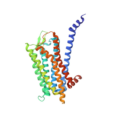
    The orexin system, which consists of the two G protein-coupled receptors OX 1 and OX 2 , activated by the neuropeptides OX-A and OX-B, is firmly established as a key regulator of behavioral arousal, sleep, and wakefulness and has been an area of intense research effort over the past two decades. X-ray structures of the receptors in complex with 10 new antagonist ligands from diverse chemotypes are presented, which complement the existing structural information for the system and highlight the critical importance of lipophilic hotspots and water molecules for these peptidergic GPCR targets. Learnings from the structural information regarding the utility of pharmacophore models and how selectivity between OX 1 and OX 2 can be achieved are discussed.


  • Organizational Affiliation

    Sosei Heptares , Steinmetz Building, Granta Park , Cambridge CB21 6DG , U.K.


Macromolecules
Find similar proteins by:  (by identity cutoff)  |  3D Structure
Entity ID: 1
MoleculeChains Sequence LengthOrganismDetailsImage
Orexin receptor type 1
A, B
368Homo sapiensMutation(s): 11 
Gene Names: HCRTR1
Membrane Entity: Yes 
UniProt & NIH Common Fund Data Resources
Find proteins for O43613 (Homo sapiens)
Explore O43613 
Go to UniProtKB:  O43613
PHAROS:  O43613
GTEx:  ENSG00000121764 
Entity Groups  
Sequence Clusters30% Identity50% Identity70% Identity90% Identity95% Identity100% Identity
UniProt GroupO43613
Sequence Annotations
Expand
  • Reference Sequence
Small Molecules
Ligands 5 Unique
IDChains Name / Formula / InChI Key2D Diagram3D Interactions
PGW
Query on PGW

Download Ideal Coordinates CCD File 
F [auth A],
S [auth B]
(1R)-2-{[(S)-{[(2S)-2,3-dihydroxypropyl]oxy}(hydroxy)phosphoryl]oxy}-1-[(hexadecanoyloxy)methyl]ethyl (9Z)-octadec-9-enoate
C40 H77 O10 P
PAZGBAOHGQRCBP-HGWHEPCSSA-N
NV8 (Subject of Investigation/LOI)
Query on NV8

Download Ideal Coordinates CCD File 
C [auth A],
P [auth B]
2-[1-(phenylsulfonyl)-1,8-diazaspiro[4.5]decan-8-yl]-1,3-benzoxazole
C21 H23 N3 O3 S
KIKWLYIDCILHAP-UHFFFAOYSA-N
SOG
Query on SOG

Download Ideal Coordinates CCD File 
G [auth A]
H [auth A]
I [auth A]
J [auth A]
K [auth A]
G [auth A],
H [auth A],
I [auth A],
J [auth A],
K [auth A],
L [auth A],
M [auth A],
N [auth A],
T [auth B],
U [auth B],
V [auth B],
W [auth B],
X [auth B]
octyl 1-thio-beta-D-glucopyranoside
C14 H28 O5 S
CGVLVOOFCGWBCS-RGDJUOJXSA-N
SO4
Query on SO4

Download Ideal Coordinates CCD File 
D [auth A],
E [auth A],
Q [auth B],
R [auth B]
SULFATE ION
O4 S
QAOWNCQODCNURD-UHFFFAOYSA-L
NA
Query on NA

Download Ideal Coordinates CCD File 
O [auth A]SODIUM ION
Na
FKNQFGJONOIPTF-UHFFFAOYSA-N
Experimental Data & Validation

Experimental Data

  • Method: X-RAY DIFFRACTION
  • Resolution: 2.30 Å
  • R-Value Free: 0.236 
  • R-Value Work: 0.209 
  • R-Value Observed: 0.210 
  • Space Group: P 1 21 1
Unit Cell:
Length ( Å )Angle ( ˚ )
a = 59.339α = 90
b = 145.892β = 112.33
c = 71.341γ = 90
Software Package:
Software NamePurpose
PHENIXrefinement
PDB_EXTRACTdata extraction
XDSdata reduction
STARANISOdata scaling
PHASERphasing

Structure Validation

View Full Validation Report


Ligand Structure Quality Assessment 


Entry History & Funding Information

Deposition Data


Funding OrganizationLocationGrant Number
National Institutes of Health/National Institute on Drug Abuse (NIH/NIDA)United StatesR01DA039553

Revision History  (Full details and data files)

  • Version 1.0: 2020-01-01
    Type: Initial release
  • Version 1.1: 2020-01-29
    Changes: Database references
  • Version 1.2: 2020-03-11
    Changes: Database references
  • Version 1.3: 2020-07-29
    Type: Remediation
    Reason: Carbohydrate remediation
    Changes: Derived calculations, Structure summary
  • Version 1.4: 2024-01-24
    Changes: Data collection, Database references, Refinement description, Structure summary