6SGF

Molecular insight into a new low affinity xylan binding module CBM86, from the xylanolytic gut symbiont Roseburia intestinalis.


Experimental Data Snapshot

  • Method: X-RAY DIFFRACTION
  • Resolution: 1.76 Å
  • R-Value Free: 0.224 
  • R-Value Work: 0.179 
  • R-Value Observed: 0.181 

wwPDB Validation   3D Report Full Report


Ligand Structure Quality Assessment 


This is version 2.0 of the entry. See complete history


Literature

Molecular insight into a new low-affinity xylan binding module from the xylanolytic gut symbiont Roseburia intestinalis.

Leth, M.L.Ejby, M.Madland, E.Kitaoku, Y.Slotboom, D.J.Guskov, A.Aachmann, F.L.Abou Hachem, M.

(2020) FEBS J 287: 2105-2117

  • DOI: https://doi.org/10.1111/febs.15117
  • Primary Citation of Related Structures:  
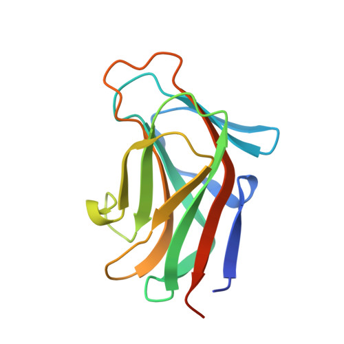
    6SGF

  • PubMed Abstract: 

    Efficient capture of glycans, the prime metabolic resources in the human gut, confers a key competitive advantage for gut microbiota members equipped with extracellular glycoside hydrolases (GHs) to target these substrates. The association of glycans to the bacterial cell surface is typically mediated by carbohydrate binding modules (CBMs). Here, we report the structure of RiCBM86 appended to a GH family 10 xylanase from Roseburia intestinalis. This CBM represents a new family of xylan binding CBMs present in xylanases from abundant and prevalent healthy human gut Clostridiales. RiCBM86 adopts a canonical β-sandwich fold, but shows structural divergence from known CBMs. The structure of RiCBM86 has been determined with a bound xylohexaose, which revealed an open and shallow binding site. RiCBM86 recognizes only a single xylosyl ring with direct hydrogen bonds. This mode of recognition is unprecedented amongst previously reported xylan binding type-B CBMs that display more extensive hydrogen-bonding patterns to their ligands or employ Ca 2+ to mediate ligand-binding. The architecture of RiCBM86 is consistent with an atypically low binding affinity (K about 0.5 mm for xylohexaose) compared to most xylan binding CBMs. Analyses using NMR spectroscopy corroborated the observations from the complex structure and the preference of RiCBM86 to arabinoxylan over glucuronoxylan, consistent with the largely negatively charged surface flanking the binding site. Mutational analysis and affinity electrophoresis established the importance of key binding residues, which are conserved in the family. This study provides novel insight into the structural features that shape low-affinity CBMs that mediate extended bacterial glycan capture in the human gut niche. DATABASES: Structural data are available in the protein data bank database under the accession number 6SGF. Sequence data are available in the GenBank database under the accession number EEV01588.1. The assignment of the Roseburia intestinalis xylan binding module into the CBM86 new family is available in the CAZy database (http://www.cazy.org/CBM86.html).


  • Organizational Affiliation

    Department of Biotechnology and Biomedicine, Technical University of Denmark, Lyngby, Denmark.


Macromolecules
Find similar proteins by:  (by identity cutoff)  |  3D Structure
Entity ID: 1
MoleculeChains Sequence LengthOrganismDetailsImage
Beta-xylanase
A, B, C, D, E
A, B, C, D, E, F
141Roseburia intestinalis L1-82Mutation(s): 0 
Gene Names: ROSINTL182_06494
EC: 3.2.1.8
UniProt
Find proteins for C7G9B5 (Roseburia intestinalis L1-82)
Explore C7G9B5 
Go to UniProtKB:  C7G9B5
Entity Groups  
Sequence Clusters30% Identity50% Identity70% Identity90% Identity95% Identity100% Identity
UniProt GroupC7G9B5
Sequence Annotations
Expand
  • Reference Sequence
Oligosaccharides

Help

Entity ID: 2
MoleculeChains Length2D Diagram Glycosylation3D Interactions
beta-D-xylopyranose-(1-4)-beta-D-xylopyranose-(1-4)-beta-D-xylopyranose
G
3N/A
Glycosylation Resources
GlyTouCan:  G87429QT
GlyCosmos:  G87429QT
Entity ID: 3
MoleculeChains Length2D Diagram Glycosylation3D Interactions
beta-D-xylopyranose-(1-4)-beta-D-xylopyranose-(1-4)-beta-D-xylopyranose-(1-4)-beta-D-xylopyranose
H, I, J, K, L
4N/A
Glycosylation Resources
GlyTouCan:  G60650FG
GlyCosmos:  G60650FG
Small Molecules
Ligands 3 Unique
IDChains Name / Formula / InChI Key2D Diagram3D Interactions
XYP (Subject of Investigation/LOI)
Query on XYP

Download Ideal Coordinates CCD File 
M [auth A]beta-D-xylopyranose
C5 H10 O5
SRBFZHDQGSBBOR-KKQCNMDGSA-N
CD
Query on CD

Download Ideal Coordinates CCD File 
BA [auth D]
CA [auth E]
N [auth A]
O [auth A]
P [auth A]
BA [auth D],
CA [auth E],
N [auth A],
O [auth A],
P [auth A],
S [auth B],
T [auth B],
V [auth C],
W [auth C],
X [auth C]
CADMIUM ION
Cd
WLZRMCYVCSSEQC-UHFFFAOYSA-N
CL
Query on CL

Download Ideal Coordinates CCD File 
AA [auth C]
DA [auth E]
EA [auth F]
Q [auth A]
R [auth A]
AA [auth C],
DA [auth E],
EA [auth F],
Q [auth A],
R [auth A],
U [auth B],
Y [auth C],
Z [auth C]
CHLORIDE ION
Cl
VEXZGXHMUGYJMC-UHFFFAOYSA-M
Biologically Interesting Molecules (External Reference) 1 Unique
Experimental Data & Validation

Experimental Data

  • Method: X-RAY DIFFRACTION
  • Resolution: 1.76 Å
  • R-Value Free: 0.224 
  • R-Value Work: 0.179 
  • R-Value Observed: 0.181 
  • Space Group: P 65
Unit Cell:
Length ( Å )Angle ( ˚ )
a = 141.87α = 90
b = 141.87β = 90
c = 60.6γ = 120
Software Package:
Software NamePurpose
PHENIXrefinement
SCALAdata scaling
PDB_EXTRACTdata extraction
XDSdata reduction
PHENIXphasing

Structure Validation

View Full Validation Report


Ligand Structure Quality Assessment 


Entry History & Funding Information

Deposition Data


Funding OrganizationLocationGrant Number
Danish Council for Independent ResearchDenmark4002-00297B

Revision History  (Full details and data files)

  • Version 1.0: 2019-11-20
    Type: Initial release
  • Version 1.1: 2020-06-03
    Changes: Data collection, Database references
  • Version 2.0: 2020-07-29
    Type: Remediation
    Reason: Carbohydrate remediation
    Changes: Advisory, Atomic model, Data collection, Derived calculations, Structure summary