6S8Y

Crystal structure of cytochrome c in complex with a sulfonated quinoline-derived foldamer


Experimental Data Snapshot

  • Method: X-RAY DIFFRACTION
  • Resolution: 2.09 Å
  • R-Value Free: 0.267 
  • R-Value Work: 0.225 
  • R-Value Observed: 0.227 

wwPDB Validation   3D Report Full Report


Ligand Structure Quality Assessment 


This is version 1.1 of the entry. See complete history


Literature

Crystal structure of a protein-aromatic foldamer composite: macromolecular chiral resolution.

Alex, J.M.Corvaglia, V.Hu, X.Engilberge, S.Huc, I.Crowley, P.B.

(2019) Chem Commun (Camb) 55: 11087-11090

  • DOI: https://doi.org/10.1039/c9cc05330a
  • Primary Citation of Related Structures:  
    6S8Y

  • PubMed Abstract: 

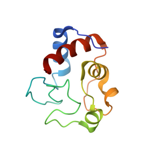
    Co-crystallization of a 2 kDa tether-free sulfonated foldamer and the 13 kDa lysine-rich cytochrome c yielded a remarkable biohybrid assembly with chiral resolution of the foldamer helix handedness. In the crystal a ∼5 nm foldamer stack was surrounded by eight molecules of protein. NMR and CD experiments suggest interesting differences in the solution state recognition processes.


  • Organizational Affiliation

    School of Chemistry, National University of Ireland, University Road, Galway, H91 TK33, Ireland.


Macromolecules
Find similar proteins by:  (by identity cutoff)  |  3D Structure
Entity ID: 1
MoleculeChains Sequence LengthOrganismDetailsImage
Cytochrome c iso-1107Saccharomyces cerevisiaeMutation(s): 0 
Gene Names: CYC1YJR048WJ1653
UniProt
Find proteins for P00044 (Saccharomyces cerevisiae (strain ATCC 204508 / S288c))
Explore P00044 
Go to UniProtKB:  P00044
Entity Groups  
Sequence Clusters30% Identity50% Identity70% Identity90% Identity95% Identity100% Identity
UniProt GroupP00044
Sequence Annotations
Expand
  • Reference Sequence
Small Molecules
Ligands 4 Unique
IDChains Name / Formula / InChI Key2D Diagram3D Interactions
L0T (Subject of Investigation/LOI)
Query on L0T

Download Ideal Coordinates CCD File 
G [auth A],
H [auth A]
8-acetamido-2-[[2-[[2-[[2-[[2-[[2-[[2-[(2-carboxy-4-sulfonato-quinolin-8-yl)carbamoyl]-4-sulfonato-quinolin-8-yl]carbamoyl]-4-sulfonato-quinolin-8-yl]carbamoyl]-4-sulfonato-quinolin-8-yl]carbamoyl]-4-sulfonato-quinolin-8-yl]carbamoyl]-4-sulfonato-quinolin-8-yl]carbamoyl]-4-sulfonato-quinolin-8-yl]carbamoyl]quinoline-4-sulfonate
C82 H44 N16 O34 S8
IHZXKZSCEKIUSH-UHFFFAOYSA-F
HEC
Query on HEC

Download Ideal Coordinates CCD File 
B [auth A]HEME C
C34 H34 Fe N4 O4
HXQIYSLZKNYNMH-LJNAALQVSA-N
ZN
Query on ZN

Download Ideal Coordinates CCD File 
C [auth A],
D [auth A],
I [auth A]
ZINC ION
Zn
PTFCDOFLOPIGGS-UHFFFAOYSA-N
ACT
Query on ACT

Download Ideal Coordinates CCD File 
E [auth A],
F [auth A]
ACETATE ION
C2 H3 O2
QTBSBXVTEAMEQO-UHFFFAOYSA-M
Experimental Data & Validation

Experimental Data

  • Method: X-RAY DIFFRACTION
  • Resolution: 2.09 Å
  • R-Value Free: 0.267 
  • R-Value Work: 0.225 
  • R-Value Observed: 0.227 
  • Space Group: P 32 2 1
Unit Cell:
Length ( Å )Angle ( ˚ )
a = 39.138α = 90
b = 39.138β = 90
c = 187.366γ = 120
Software Package:
Software NamePurpose
BUSTERrefinement
PDB_EXTRACTdata extraction
XDSdata reduction
Aimlessdata scaling
MOLREPphasing

Structure Validation

View Full Validation Report


Ligand Structure Quality Assessment 


Entry History & Funding Information

Deposition Data


Funding OrganizationLocationGrant Number
Science Foundation IrelandIreland13/CDA/2168

Revision History  (Full details and data files)

  • Version 1.0: 2019-09-25
    Type: Initial release
  • Version 1.1: 2024-01-24
    Changes: Data collection, Database references, Derived calculations, Refinement description