6S8V

Structure of the high affinity Anticalin P3D11 in complex with the human CD98 heavy chain ectodomain


Experimental Data Snapshot

  • Method: X-RAY DIFFRACTION
  • Resolution: 1.80 Å
  • R-Value Free: 0.226 
  • R-Value Work: 0.184 
  • R-Value Observed: 0.185 

wwPDB Validation   3D Report Full Report


This is version 1.2 of the entry. See complete history


Literature

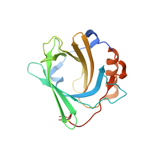
Development of a high affinity Anticalin®directed against human CD98hc for theranostic applications.

Deuschle, F.C.Morath, V.Schiefner, A.Brandt, C.Ballke, S.Reder, S.Steiger, K.Schwaiger, M.Weber, W.Skerra, A.

(2020) Theranostics 10: 2172-2187

  • DOI: https://doi.org/10.7150/thno.38968
  • Primary Citation of Related Structures:  
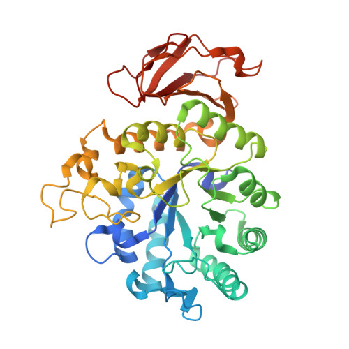
    6S8V

  • PubMed Abstract: 

    Enhanced amino acid supply and dysregulated integrin signaling constitute two hallmarks of cancer and are pivotal for metastatic transformation of cells. In line with its function at the crossroads of both processes, overexpression of CD98hc is clinically observed in various cancer malignancies, thus rendering it a promising tumor target. Methods : We describe the development of Anticalin proteins based on the lipocalin 2 (Lcn2) scaffold against the human CD98hc ectodomain (hCD98hcED) using directed evolution and protein design. X-ray structural analysis was performed to identify the epitope recognized by the lead Anticalin candidate. The Anticalin - with a tuned plasma half-life using PASylation ® technology - was labeled with 89 Zr and investigated by positron emission tomography (PET) of CD98-positive tumor xenograft mice. Results : The Anticalin P3D11 binds CD98hc with picomolar affinity and recognizes a protruding loop structure surrounded by several glycosylation sites within the solvent exposed membrane-distal part of the hCD98hcED. In vitro studies revealed specific binding activity of the Anticalin towards various CD98hc-expressing human tumor cell lines, suggesting broader applicability in cancer research. PET/CT imaging of mice bearing human prostate carcinoma xenografts using the optimized and 89 Zr-labeled Anticalin demonstrated strong and specific tracer accumulation (8.6 ± 1.1 %ID/g) as well as a favorable tumor-to-blood ratio of 11.8. Conclusion : Our findings provide a first proof of concept to exploit CD98hc for non-invasive biomedical imaging. The novel Anticalin-based αhCD98hc radiopharmaceutical constitutes a promising tool for preclinical and, potentially, clinical applications in oncology.


  • Organizational Affiliation

    Lehrstuhl für Biologische Chemie, Technische Universität München, 85354 Freising, Germany.


Macromolecules
Find similar proteins by:  (by identity cutoff)  |  3D Structure
Entity ID: 1
MoleculeChains Sequence LengthOrganismDetailsImage
Neutrophil gelatinase-associated lipocalin
A, C
186Homo sapiensMutation(s): 21 
Gene Names: LCN2HNLNGAL
UniProt & NIH Common Fund Data Resources
Find proteins for P80188 (Homo sapiens)
Explore P80188 
Go to UniProtKB:  P80188
PHAROS:  P80188
GTEx:  ENSG00000148346 
Entity Groups  
Sequence Clusters30% Identity50% Identity70% Identity90% Identity95% Identity100% Identity
UniProt GroupP80188
Sequence Annotations
Expand
  • Reference Sequence
Find similar proteins by:  (by identity cutoff)  |  3D Structure
Entity ID: 2
MoleculeChains Sequence LengthOrganismDetailsImage
4F2 cell-surface antigen heavy chain
B, D
431Homo sapiensMutation(s): 0 
Gene Names: SLC3A2MDU1
UniProt & NIH Common Fund Data Resources
Find proteins for P08195 (Homo sapiens)
Explore P08195 
Go to UniProtKB:  P08195
PHAROS:  P08195
GTEx:  ENSG00000168003 
Entity Groups  
Sequence Clusters30% Identity50% Identity70% Identity90% Identity95% Identity100% Identity
UniProt GroupP08195
Sequence Annotations
Expand
  • Reference Sequence
Experimental Data & Validation

Experimental Data

  • Method: X-RAY DIFFRACTION
  • Resolution: 1.80 Å
  • R-Value Free: 0.226 
  • R-Value Work: 0.184 
  • R-Value Observed: 0.185 
  • Space Group: C 1 2 1
Unit Cell:
Length ( Å )Angle ( ˚ )
a = 202.809α = 90
b = 46.052β = 106.64
c = 137.092γ = 90
Software Package:
Software NamePurpose
XSCALEdata scaling
REFMACrefinement
PDB_EXTRACTdata extraction
XDSdata reduction
PHASERphasing

Structure Validation

View Full Validation Report


Entry History & Funding Information

Deposition Data


Funding OrganizationLocationGrant Number
German Research FoundationGermanyCollaborative Research Centre 824 project A8

Revision History  (Full details and data files)

  • Version 1.0: 2020-03-04
    Type: Initial release
  • Version 1.1: 2020-03-11
    Changes: Data collection
  • Version 1.2: 2024-01-24
    Changes: Data collection, Database references, Refinement description