6RQX

High-resolution crystal structure of ERAP1 in complex with 10mer phosphinic peptide


Experimental Data Snapshot

  • Method: X-RAY DIFFRACTION
  • Resolution: 1.68 Å
  • R-Value Free: 0.209 
  • R-Value Work: 0.176 
  • R-Value Observed: 0.176 

wwPDB Validation   3D Report Full Report


Ligand Structure Quality Assessment 


This is version 2.1 of the entry. See complete history


Literature

Mechanism for antigenic peptide selection by endoplasmic reticulum aminopeptidase 1.

Giastas, P.Mpakali, A.Papakyriakou, A.Lelis, A.Kokkala, P.Neu, M.Rowland, P.Liddle, J.Georgiadis, D.Stratikos, E.

(2019) Proc Natl Acad Sci U S A 

  • DOI: https://doi.org/10.1073/pnas.1912070116
  • Primary Citation of Related Structures:  
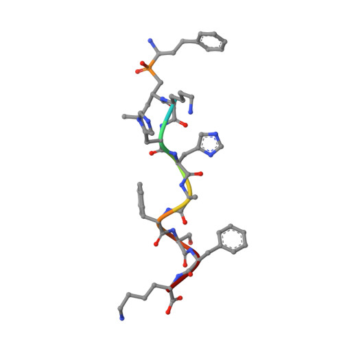
    6RQX, 6RYF

  • PubMed Abstract: 

    Endoplasmic reticulum aminopeptidase 1 (ERAP1) is an intracellular enzyme that optimizes the peptide cargo of major histocompatibility class I (MHC-I) molecules and regulates adaptive immunity. It has unusual substrate selectivity for length and sequence, resulting in poorly understood effects on the cellular immunopeptidome. To understand substrate selection by ERAP1, we solved 2 crystal structures of the enzyme with bound transition-state pseudopeptide analogs at 1.68 Å and 1.72 Å. Both peptides have their N terminus bound at the active site and extend away along a large internal cavity, interacting with shallow pockets that can influence selectivity. The longer peptide is disordered through the central region of the cavity and has its C terminus bound in an allosteric pocket of domain IV that features a carboxypeptidase-like structural motif. These structures, along with enzymatic and computational analyses, explain how ERAP1 can select peptides based on length while retaining the broad sequence-specificity necessary for its biological function.


  • Organizational Affiliation

    Institute of Nuclear & Radiological Sciences and Technology, Energy & Safety, National Centre for Scientific Research Demokritos, Agia Paraskevi, Athens 15341, Greece.


Macromolecules
Find similar proteins by:  (by identity cutoff)  |  3D Structure
Entity ID: 1
MoleculeChains Sequence LengthOrganismDetailsImage
Endoplasmic reticulum aminopeptidase 1938Homo sapiensMutation(s): 0 
Gene Names: ERAP1APPILSARTS1KIAA0525UNQ584/PRO1154
EC: 3.4.11
UniProt & NIH Common Fund Data Resources
Find proteins for Q9NZ08 (Homo sapiens)
Explore Q9NZ08 
Go to UniProtKB:  Q9NZ08
PHAROS:  Q9NZ08
GTEx:  ENSG00000164307 
Entity Groups  
Sequence Clusters30% Identity50% Identity70% Identity90% Identity95% Identity100% Identity
UniProt GroupQ9NZ08
Sequence Annotations
Expand
  • Reference Sequence

Find similar proteins by:  Sequence   |   3D Structure  

Entity ID: 2
MoleculeChains Sequence LengthOrganismDetailsImage
PSE-LYS-HIS-HIS-ALA-PHE-SER-PHE-LYS9Homo sapiensMutation(s): 0 
Sequence Annotations
Expand
  • Reference Sequence
Oligosaccharides

Help

Entity ID: 3
MoleculeChains Length2D Diagram Glycosylation3D Interactions
alpha-L-fucopyranose-(1-3)-[2-acetamido-2-deoxy-beta-D-glucopyranose-(1-4)]2-acetamido-2-deoxy-beta-D-glucopyranose
C
3N-Glycosylation
Glycosylation Resources
GlyTouCan:  G11971MR
GlyCosmos:  G11971MR
GlyGen:  G11971MR
Entity ID: 4
MoleculeChains Length2D Diagram Glycosylation3D Interactions
alpha-D-mannopyranose-(1-3)-beta-D-mannopyranose-(1-4)-2-acetamido-2-deoxy-beta-D-glucopyranose-(1-4)-2-acetamido-2-deoxy-beta-D-glucopyranose
D
4N-Glycosylation
Glycosylation Resources
GlyTouCan:  G81315DD
GlyCosmos:  G81315DD
GlyGen:  G81315DD
Entity ID: 5
MoleculeChains Length2D Diagram Glycosylation3D Interactions
2-acetamido-2-deoxy-beta-D-glucopyranose-(1-4)-2-acetamido-2-deoxy-beta-D-glucopyranose
E
2N-Glycosylation
Glycosylation Resources
GlyTouCan:  G42666HT
GlyCosmos:  G42666HT
GlyGen:  G42666HT
Small Molecules
Ligands 6 Unique
IDChains Name / Formula / InChI Key2D Diagram3D Interactions
NAG
Query on NAG

Download Ideal Coordinates CCD File 
F [auth A]2-acetamido-2-deoxy-beta-D-glucopyranose
C8 H15 N O6
OVRNDRQMDRJTHS-FMDGEEDCSA-N
PGE
Query on PGE

Download Ideal Coordinates CCD File 
SA [auth A],
TA [auth A],
UA [auth A]
TRIETHYLENE GLYCOL
C6 H14 O4
ZIBGPFATKBEMQZ-UHFFFAOYSA-N
ZN
Query on ZN

Download Ideal Coordinates CCD File 
G [auth A]ZINC ION
Zn
PTFCDOFLOPIGGS-UHFFFAOYSA-N
EDO
Query on EDO

Download Ideal Coordinates CCD File 
AA [auth A]
BA [auth A]
CA [auth A]
DA [auth A]
EA [auth A]
AA [auth A],
BA [auth A],
CA [auth A],
DA [auth A],
EA [auth A],
FA [auth A],
GA [auth A],
HA [auth A],
IA [auth A],
JA [auth A],
KA [auth A],
LA [auth A],
MA [auth A],
NA [auth A],
S [auth A],
T [auth A],
U [auth A],
V [auth A],
W [auth A],
X [auth A],
Y [auth A],
Z [auth A]
1,2-ETHANEDIOL
C2 H6 O2
LYCAIKOWRPUZTN-UHFFFAOYSA-N
SCN
Query on SCN

Download Ideal Coordinates CCD File 
H [auth A]
I [auth A]
J [auth A]
K [auth A]
L [auth A]
H [auth A],
I [auth A],
J [auth A],
K [auth A],
L [auth A],
M [auth A],
N [auth A],
O [auth A],
P [auth A],
Q [auth A],
R [auth A]
THIOCYANATE ION
C N S
ZMZDMBWJUHKJPS-UHFFFAOYSA-M
NA
Query on NA

Download Ideal Coordinates CCD File 
OA [auth A],
PA [auth A],
QA [auth A],
RA [auth A]
SODIUM ION
Na
FKNQFGJONOIPTF-UHFFFAOYSA-N
Experimental Data & Validation

Experimental Data

  • Method: X-RAY DIFFRACTION
  • Resolution: 1.68 Å
  • R-Value Free: 0.209 
  • R-Value Work: 0.176 
  • R-Value Observed: 0.176 
  • Space Group: P 2 21 21
Unit Cell:
Length ( Å )Angle ( ˚ )
a = 58.004α = 90
b = 116.439β = 90
c = 147.363γ = 90
Software Package:
Software NamePurpose
PHENIXrefinement
XDSdata reduction
XDSdata scaling
PHASERphasing

Structure Validation

View Full Validation Report


Ligand Structure Quality Assessment 


Entry History 

Deposition Data

Revision History  (Full details and data files)

  • Version 1.0: 2019-12-18
    Type: Initial release
  • Version 1.1: 2020-07-01
    Changes: Data collection, Database references
  • Version 2.0: 2020-07-29
    Type: Remediation
    Reason: Carbohydrate remediation
    Changes: Atomic model, Data collection, Derived calculations, Structure summary
  • Version 2.1: 2024-01-24
    Changes: Advisory, Data collection, Database references, Derived calculations, Refinement description, Structure summary