6RAR

Pmar-Lig_PreS3-Mn


Experimental Data Snapshot

  • Method: X-RAY DIFFRACTION
  • Resolution: 1.78 Å
  • R-Value Free: 0.264 
  • R-Value Work: 0.233 
  • R-Value Observed: 0.234 

wwPDB Validation   3D Report Full Report


Ligand Structure Quality Assessment 


This is version 1.2 of the entry. See complete history

Re-refinement Note

A newer entry is available that reflects an alternative modeling of the original data: 6RCE


Literature

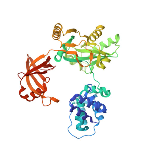
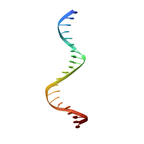
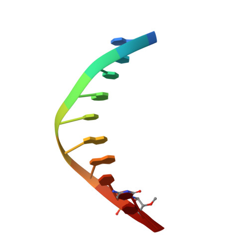
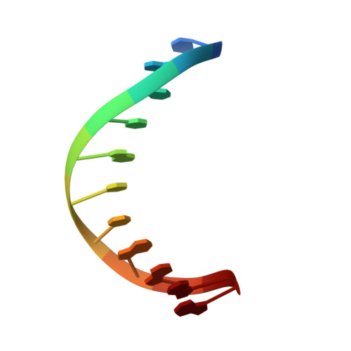
Structural intermediates of a DNA-ligase complex illuminate the role of the catalytic metal ion and mechanism of phosphodiester bond formation.

Williamson, A.Leiros, H.S.

(2019) Nucleic Acids Res 47: 7147-7162

  • DOI: https://doi.org/10.1093/nar/gkz596
  • Primary Citation of Related Structures:  
    6RAR, 6RAS, 6RAU, 6RCE

  • PubMed Abstract: 

    DNA ligases join adjacent 5' phosphate (5'P) and 3' hydroxyl (3'OH) termini of double-stranded DNA via a three-step mechanism requiring a nucleotide cofactor and divalent metal ion. Although considerable structural detail is available for the first two steps, less is known about step 3 where the DNA-backbone is joined or about the cation role at this step. We have captured high-resolution structures of an adenosine triphosphate (ATP)-dependent DNA ligase from Prochlorococcus marinus including a Mn-bound pre-ternary ligase-DNA complex poised for phosphodiester bond formation, and a post-ternary intermediate retaining product DNA and partially occupied AMP in the active site. The pre-ternary structure unambiguously identifies the binding site of the catalytic metal ion and confirms both its role in activating the 3'OH terminus for nucleophilic attack on the 5'P group and stabilizing the pentavalent transition state. The post-ternary structure indicates that DNA distortion and most enzyme-AMP contacts remain after phosphodiester bond formation, implying loss of covalent linkage to the DNA drives release of AMP, rather than active site rearrangement. Additionally, comparisons of this cyanobacterial DNA ligase with homologs from bacteria and bacteriophage pose interesting questions about the structural origin of double-strand break joining activity and the evolution of these ATP-dependent DNA ligase enzymes.


  • Organizational Affiliation

    Department of Chemistry, UiT The Arctic University of Norway, Tromsø, N-9037, Norway.


Macromolecules

Find similar proteins by:  (by identity cutoff)  |  3D Structure
Entity ID: 4
MoleculeChains Sequence LengthOrganismDetailsImage
ATP-dependent DNA ligaseD [auth I]432Prochlorococcus marinus str. MIT 9302Mutation(s): 0 
Gene Names: EU96_0746
UniProt
Find proteins for A0A0A2ACP7 (Prochlorococcus marinus str. MIT 9302)
Explore A0A0A2ACP7 
Go to UniProtKB:  A0A0A2ACP7
Entity Groups  
Sequence Clusters30% Identity50% Identity70% Identity90% Identity95% Identity100% Identity
UniProt GroupA0A0A2ACP7
Sequence Annotations
Expand
  • Reference Sequence

Find similar nucleic acids by:  Sequence   |   3D Structure  

Entity ID: 1
MoleculeChains LengthOrganismImage
DNA (5'-D(*TP*TP*CP*CP*GP*AP*CP*AP*GP*TP*GP*GP*GP*GP*TP*CP*GP*CP*AP*AP*T)-3')A [auth B]21synthetic construct
Sequence Annotations
Expand
  • Reference Sequence

Find similar nucleic acids by:  Sequence   |   3D Structure  

Entity ID: 2
MoleculeChains LengthOrganismImage
DNA/RNA (5'-D(*AP*TP*TP*GP*CP*GP*AP*C)-R(P*(OMC))-D(P*C)-3')B [auth C]10synthetic construct
Sequence Annotations
Expand
  • Reference Sequence

Find similar nucleic acids by:  Sequence   |   3D Structure  

Entity ID: 3
MoleculeChains LengthOrganismImage
DNA (5'-D(P*CP*AP*CP*TP*AP*TP*CP*GP*GP*AP*A)-3')C [auth D]11synthetic construct
Sequence Annotations
Expand
  • Reference Sequence
Experimental Data & Validation

Experimental Data

  • Method: X-RAY DIFFRACTION
  • Resolution: 1.78 Å
  • R-Value Free: 0.264 
  • R-Value Work: 0.233 
  • R-Value Observed: 0.234 
  • Space Group: P 1 21 1
Unit Cell:
Length ( Å )Angle ( ˚ )
a = 67.304α = 90
b = 103.315β = 90.79
c = 68.543γ = 90
Software Package:
Software NamePurpose
PHENIXrefinement
XDSdata reduction
Aimlessdata scaling
PHASERphasing

Structure Validation

View Full Validation Report


Ligand Structure Quality Assessment 


Entry History & Funding Information

Deposition Data


Funding OrganizationLocationGrant Number
Research Council of NorwayNorway244247, 2015

Revision History  (Full details and data files)

  • Version 1.0: 2019-07-10
    Type: Initial release
  • Version 1.1: 2020-01-29
    Changes: Database references
  • Version 1.2: 2024-01-24
    Changes: Data collection, Database references, Derived calculations, Refinement description