6R8R

Structure of the Wnt deacylase Notum in complex with isoquinoline 45


Experimental Data Snapshot

  • Method: X-RAY DIFFRACTION
  • Resolution: 1.27 Å
  • R-Value Free: 0.178 
  • R-Value Work: 0.148 
  • R-Value Observed: 0.149 

wwPDB Validation   3D Report Full Report


Ligand Structure Quality Assessment 


This is version 1.4 of the entry. See complete history


Literature

Discovery of 2-phenoxyacetamides as inhibitors of the Wnt-depalmitoleating enzyme NOTUM from an X-ray fragment screen.

Atkinson, B.N.Steadman, D.Zhao, Y.Sipthorp, J.Vecchia, L.Ruza, R.R.Jeganathan, F.Lines, G.Frew, S.Monaghan, A.Kjær, S.Bictash, M.Jones, E.Y.Fish, P.V.

(2019) Medchemcomm 10: 1361-1369

  • DOI: https://doi.org/10.1039/c9md00096h
  • Primary Citation of Related Structures:  
    6R8P, 6R8Q, 6R8R

  • PubMed Abstract: 

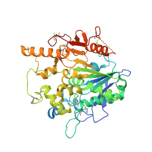
    NOTUM is a carboxylesterase that has been shown to act by mediating the O -depalmitoleoylation of Wnt proteins resulting in suppression of Wnt signaling. Here, we describe the development of NOTUM inhibitors that restore Wnt signaling for use in in vitro disease models where NOTUM over activity is an underlying cause. A crystallographic fragment screen with NOTUM identified 2-phenoxyacetamide 3 as binding in the palmitoleate pocket with modest inhibition activity (IC 50 33 μM). Optimization of hit 3 by SAR studies guided by SBDD identified indazole 38 (IC 50 0.032 μM) and isoquinoline 45 (IC 50 0.085 μM) as potent inhibitors of NOTUM. The binding of 45 to NOTUM was rationalized through an X-ray co-crystal structure determination which showed a flipped binding orientation compared to 3 . However, it was not possible to combine NOTUM inhibition activity with metabolic stability as the majority of the compounds tested were rapidly metabolized in an NADPH-independent manner.


  • Organizational Affiliation

    Alzheimer's Research UK UCL Drug Discovery Institute , University College London , Cruciform Building, Gower Street , London , WC1E 6BT , UK . Email: p.fish@ucl.ac.uk ; Tel: +44 (0)20 7679 6971.


Macromolecules
Find similar proteins by:  (by identity cutoff)  |  3D Structure
Entity ID: 1
MoleculeChains Sequence LengthOrganismDetailsImage
Palmitoleoyl-protein carboxylesterase NOTUM383Homo sapiensMutation(s): 1 
Gene Names: NOTUMOK/SW-CL.30
EC: 3.1.1.98
UniProt & NIH Common Fund Data Resources
Find proteins for Q6P988 (Homo sapiens)
Explore Q6P988 
Go to UniProtKB:  Q6P988
PHAROS:  Q6P988
GTEx:  ENSG00000185269 
Entity Groups  
Sequence Clusters30% Identity50% Identity70% Identity90% Identity95% Identity100% Identity
UniProt GroupQ6P988
Sequence Annotations
Expand
  • Reference Sequence
Small Molecules
Ligands 5 Unique
IDChains Name / Formula / InChI Key2D Diagram3D Interactions
JV8 (Subject of Investigation/LOI)
Query on JV8

Download Ideal Coordinates CCD File 
C [auth A]~{N}-isoquinolin-6-yl-2-(2-methylphenoxy)ethanamide
C18 H16 N2 O2
PEVCERQGPZRIFE-UHFFFAOYSA-N
NAG
Query on NAG

Download Ideal Coordinates CCD File 
B [auth A]2-acetamido-2-deoxy-beta-D-glucopyranose
C8 H15 N O6
OVRNDRQMDRJTHS-FMDGEEDCSA-N
SO4
Query on SO4

Download Ideal Coordinates CCD File 
D [auth A]
E [auth A]
F [auth A]
G [auth A]
H [auth A]
D [auth A],
E [auth A],
F [auth A],
G [auth A],
H [auth A],
I [auth A],
J [auth A],
K [auth A]
SULFATE ION
O4 S
QAOWNCQODCNURD-UHFFFAOYSA-L
DMS
Query on DMS

Download Ideal Coordinates CCD File 
L [auth A],
M [auth A],
N [auth A],
O [auth A],
P [auth A]
DIMETHYL SULFOXIDE
C2 H6 O S
IAZDPXIOMUYVGZ-UHFFFAOYSA-N
EDO
Query on EDO

Download Ideal Coordinates CCD File 
Q [auth A]
R [auth A]
S [auth A]
T [auth A]
U [auth A]
Q [auth A],
R [auth A],
S [auth A],
T [auth A],
U [auth A],
V [auth A],
W [auth A],
X [auth A],
Y [auth A],
Z [auth A]
1,2-ETHANEDIOL
C2 H6 O2
LYCAIKOWRPUZTN-UHFFFAOYSA-N
Binding Affinity Annotations 
IDSourceBinding Affinity
JV8 Binding MOAD:  6R8R IC50: 85 (nM) from 1 assay(s)
Experimental Data & Validation

Experimental Data

  • Method: X-RAY DIFFRACTION
  • Resolution: 1.27 Å
  • R-Value Free: 0.178 
  • R-Value Work: 0.148 
  • R-Value Observed: 0.149 
  • Space Group: P 21 21 21
Unit Cell:
Length ( Å )Angle ( ˚ )
a = 60.34α = 90
b = 71.72β = 90
c = 78.13γ = 90
Software Package:
Software NamePurpose
PHENIXrefinement
XSCALEdata scaling
PDB_EXTRACTdata extraction
XDSdata reduction
PHASERphasing

Structure Validation

View Full Validation Report


Ligand Structure Quality Assessment 


Entry History & Funding Information

Deposition Data


Funding OrganizationLocationGrant Number
Cancer Research UKUnited KingdomC375/A17721
Medical Research Council (United Kingdom)United KingdomMR/M000141/1

Revision History  (Full details and data files)

  • Version 1.0: 2019-05-08
    Type: Initial release
  • Version 1.1: 2019-07-10
    Changes: Data collection
  • Version 1.2: 2019-10-02
    Changes: Data collection, Database references
  • Version 1.3: 2020-07-29
    Type: Remediation
    Reason: Carbohydrate remediation
    Changes: Data collection, Derived calculations, Structure summary
  • Version 1.4: 2024-01-24
    Changes: Data collection, Database references, Refinement description, Structure summary