


6R35
Structure of the LecB lectin from Pseudomonas aeruginosa strain PAO1 in complex with lewis x tetrasaccharide
- PDB DOI: https://doi.org/10.2210/pdb6R35/pdb
- Classification: SUGAR BINDING PROTEIN
- Organism(s): Pseudomonas aeruginosa PAO1
- Expression System: Escherichia coli BL21(DE3)
- Mutation(s): No
- Deposited: 2019-03-19 Released: 2019-06-12
- Funding Organization(s): European Union
Experimental Data Snapshot
- Method: X-RAY DIFFRACTION
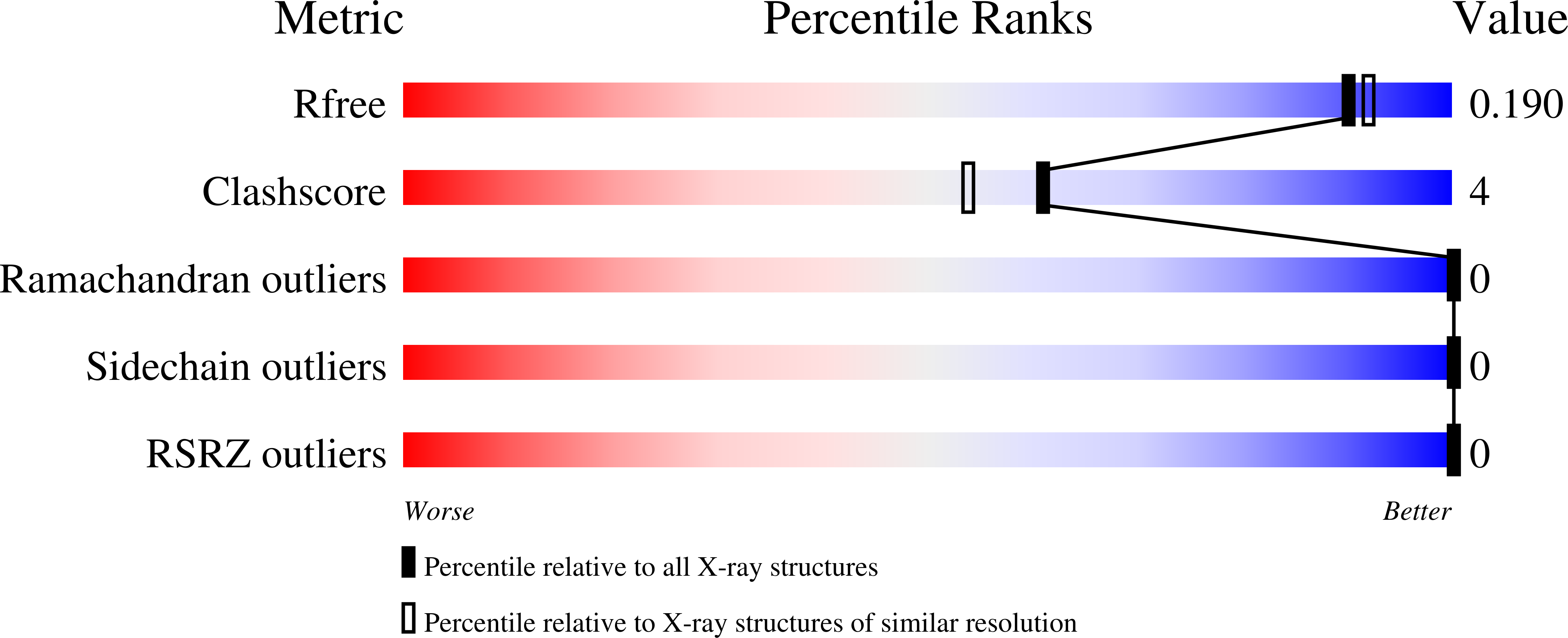
- Resolution: 1.80 Å
- R-Value Free: 0.176
- R-Value Work: 0.132
- R-Value Observed: 0.134
wwPDB Validation 3D Report Full Report
This is version 2.1 of the entry. See complete history.

Macromolecules
Find similar proteins by:
(by identity cutoff) | 3D Structure
Entity ID: 1 | |||||
|---|---|---|---|---|---|
| Molecule | Chains | Sequence Length | Organism | Details | Image |
| Fucose-binding lectin PA-IIL | A [auth D], B [auth A], C [auth B], D [auth C] | 114 | Pseudomonas aeruginosa PAO1 | Mutation(s): 0 Gene Names: lecB, PA3361 |  |
UniProt | |||||
Find proteins for Q9HYN5 (Pseudomonas aeruginosa (strain ATCC 15692 / DSM 22644 / CIP 104116 / JCM 14847 / LMG 12228 / 1C / PRS 101 / PAO1)) Explore Q9HYN5 Go to UniProtKB: Q9HYN5 | |||||
Entity Groups | |||||
| Sequence Clusters | 30% Identity50% Identity70% Identity90% Identity95% Identity100% Identity | ||||
| UniProt Group | Q9HYN5 | ||||
Sequence AnnotationsExpand | |||||
| |||||
Oligosaccharides

Small Molecules
| Ligands 3 Unique | |||||
|---|---|---|---|---|---|
| ID | Chains | Name / Formula / InChI Key | 2D Diagram | 3D Interactions | |
| SO4 Query on SO4 | K [auth D], W [auth C] | SULFATE ION O4 S QAOWNCQODCNURD-UHFFFAOYSA-L |  | ||
| EDO Query on EDO | L [auth D] P [auth A] Q [auth A] T [auth B] U [auth B] | 1,2-ETHANEDIOL C2 H6 O2 LYCAIKOWRPUZTN-UHFFFAOYSA-N |  | ||
| CA (Subject of Investigation/LOI) Query on CA | I [auth D] J [auth D] M [auth D] N [auth A] O [auth A] | CALCIUM ION Ca BHPQYMZQTOCNFJ-UHFFFAOYSA-N |  | ||
Biologically Interesting Molecules (External Reference) 1 Unique
Entity ID: 2 | |||||
|---|---|---|---|---|---|
| ID | Chains | Name | Type/Class | 2D Diagram | 3D Interactions |
| PRD_900119 Query on PRD_900119 | E, F, G, H | Lewis X antigen, beta anomer | Oligosaccharide / Antigen |  | |
Experimental Data & Validation
Experimental Data
- Method: X-RAY DIFFRACTION
- Resolution: 1.80 Å
- R-Value Free: 0.176
- R-Value Work: 0.132
- R-Value Observed: 0.134
- Space Group: P 1 21 1
Unit Cell:
| Length ( Å ) | Angle ( ˚ ) |
|---|---|
| a = 52.603 | α = 90 |
| b = 72.481 | β = 114.62 |
| c = 62.061 | γ = 90 |
| Software Name | Purpose |
|---|---|
| XDS | data reduction |
| Aimless | data scaling |
| REFMAC | refinement |
| PDB_EXTRACT | data extraction |
| PHASER | phasing |
Entry History & Funding Information
Deposition Data
- Released Date: 2019-06-12 Deposition Author(s): Lepsik, M., Sommer, R., Kuhaudomlarp, S., Lelimousin, M., Varrot, A., Titz, A., Imberty, A.
| Funding Organization | Location | Grant Number |
|---|---|---|
| European Union | France | 795605 |
Revision History (Full details and data files)
- Version 1.0: 2019-06-12
Type: Initial release - Version 1.1: 2019-09-18
Changes: Data collection, Refinement description - Version 2.0: 2020-07-29
Type: Remediation
Reason: Carbohydrate remediation
Changes: Atomic model, Data collection, Derived calculations, Structure summary - Version 2.1: 2024-01-24
Changes: Data collection, Database references, Refinement description, Structure summary
